Functional changes
Alterations in brain functioning are investigated by using imaging techniques such as functional magnetic resonance imaging and electroencephalogram, and by assessing changes in biochemicals and proteins that are known to be effected by stress. Click on the tabs below to access the information, or navigate the topics via the section on the left.
Image: ©fabioberti.it – stock.adobe.com
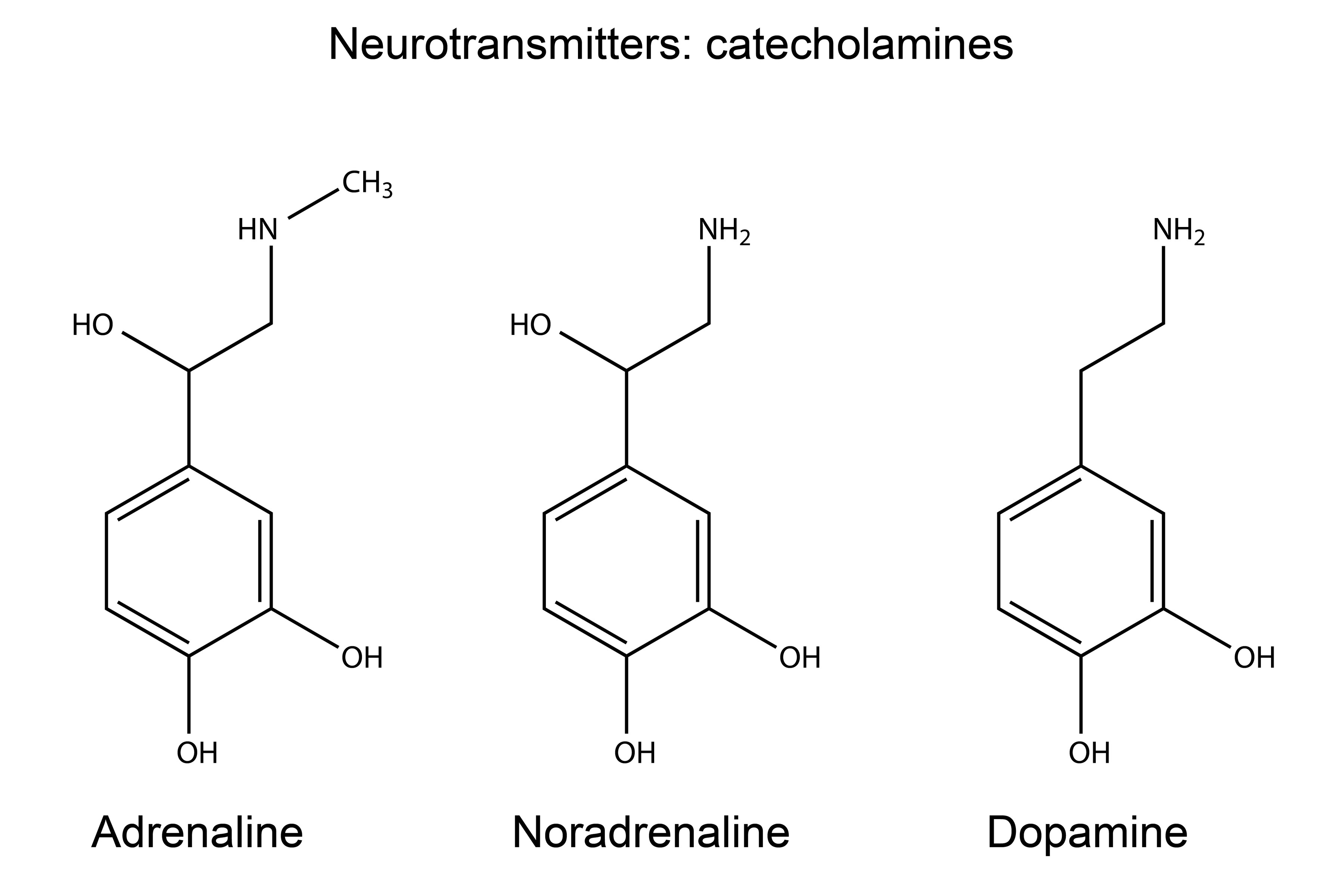
Catecholamines
What are catecholamines in PTSD? Catecholamines are a group of neurotransmitters that include dopamine, norepinephrine, also called noradrenaline, and epinephrine, also called adrenaline. The sympathetic nervous system stimulates the release of catecholamines to mediate adaptive responses to acute stress. Catecholamines are also linked to long-term memory of events that induce strong emotions, including fear. Stress-responsive neurotransmitters released during emotional arousal are thought to enhance the consolidation of fear memory. Hyperresponsiveness in the dopaminergic system is common in individuals who have been exposed to stress, which was associated with PTSD symptoms such as restlessness, nightmares, fear memory, and impulsivity. Over activation…

EEG
What is electroencephalography (EEG) and PTSD? Electroencephalography (EEG) has been used to study changes in brain functioning in people with PTSD. It uses electrodes on the scalp to measure electrical activity from the brain. Quantitative spectral EEG investigates several waveforms so the activity can be measured. However, EEG also gives rise to event related potentials (ERP). These measure the EEG activity directly evoked by a stimulus, often using cognitive or perceptual stimuli. P300, also referred to as P3, may be the ERP most suitable for the assessment of PTSD. This is because it is well documented, and with the appropriate…
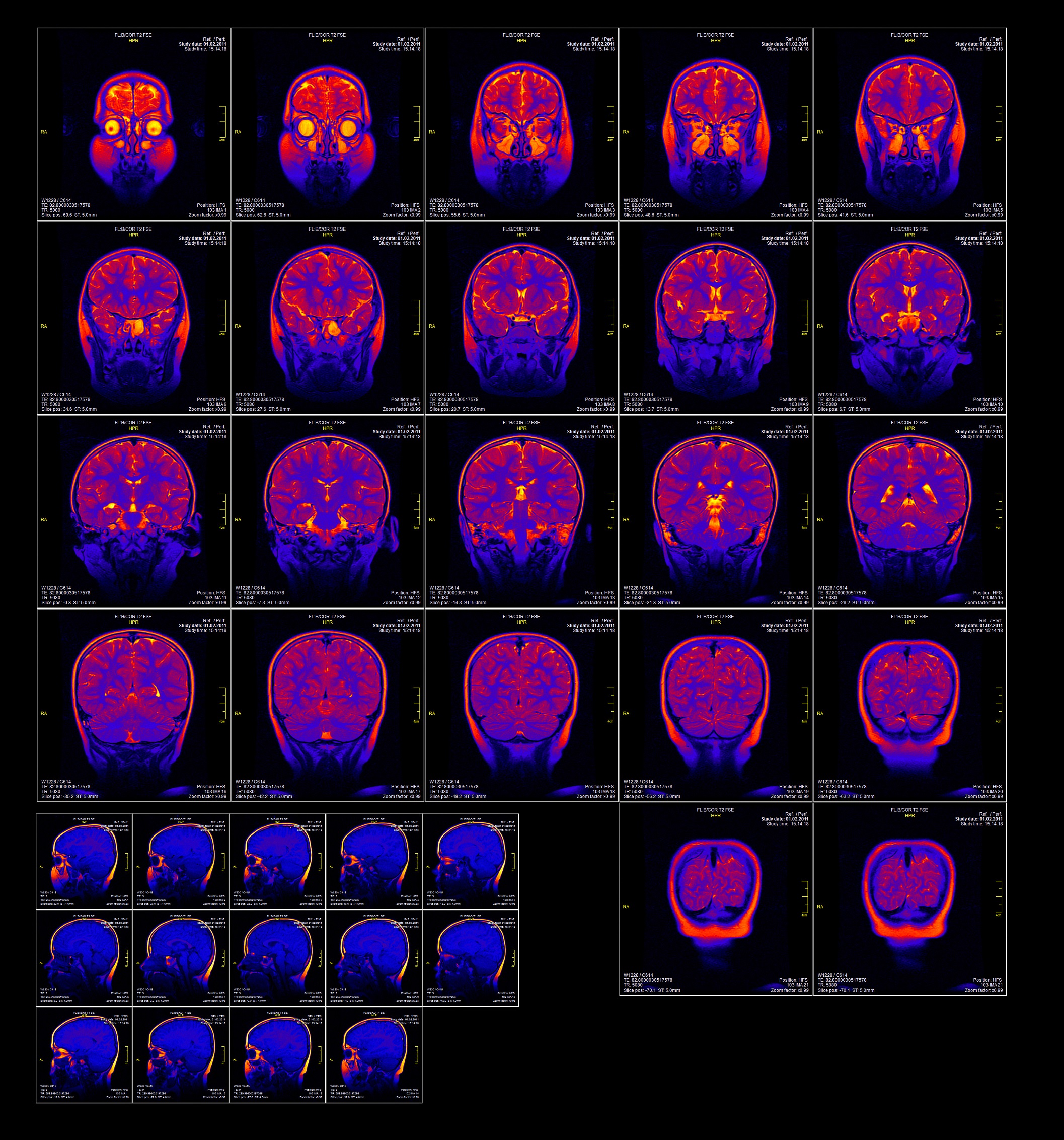
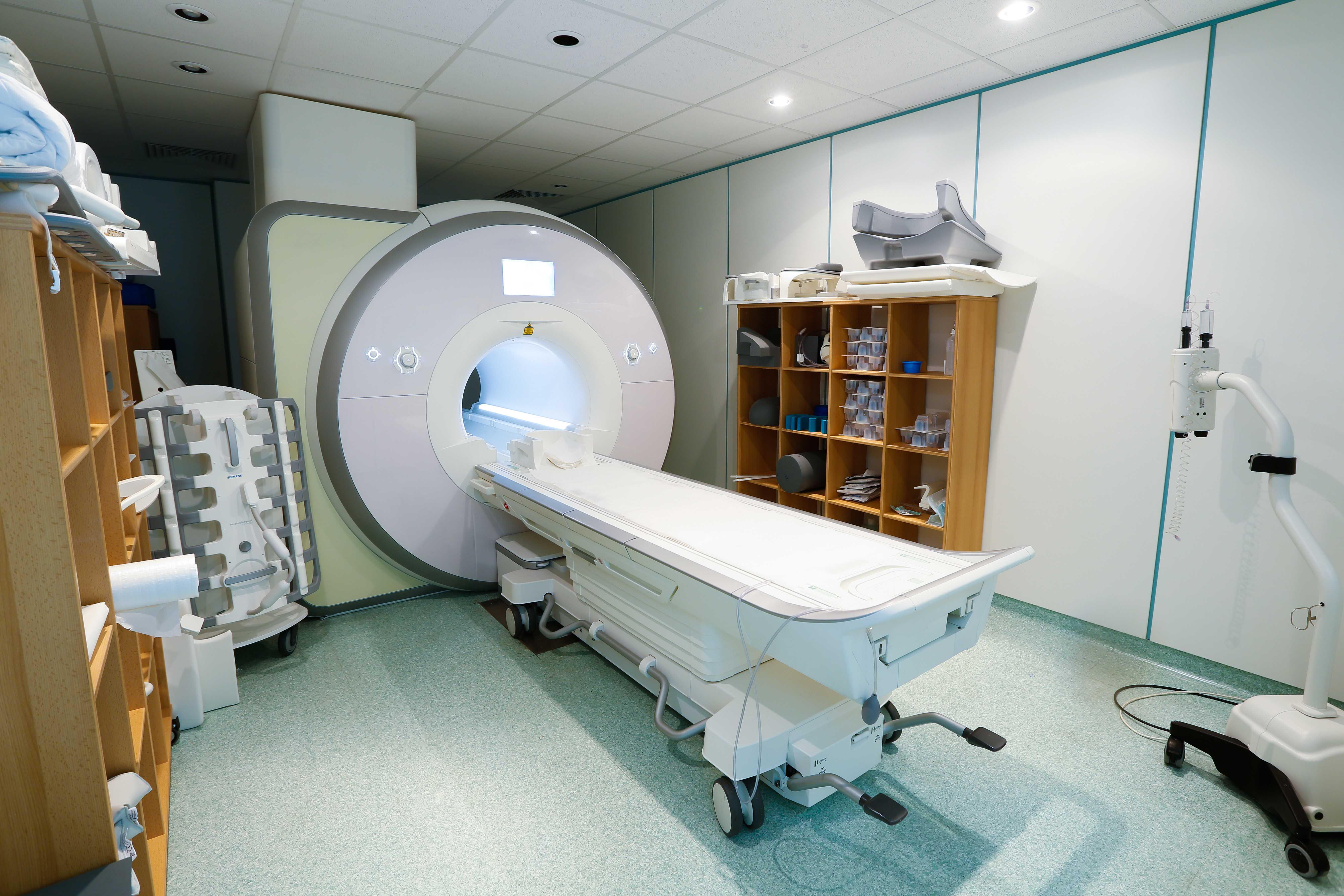
fMRI and PET
What is brain functioning in PTSD, measured with fMRI, PET and SPECT? Functional magnetic resonance imaging (fMRI) measures blood flow to determine activation and deactivation of specific brain regions. Positron emission tomography (PET) is a nuclear based imaging technique that utilises a radioactive tracer to visualise functional brain activity. The radioisotope tracers are coupled with a biological molecule such as glucose, which is used during cellular metabolism and can be used to highlight areas with changes in metabolic activity. Single-photon emission computed tomography (SPECT) offers more limited spatial and temporal resolution than PET but is less expensive as it does…
GABA
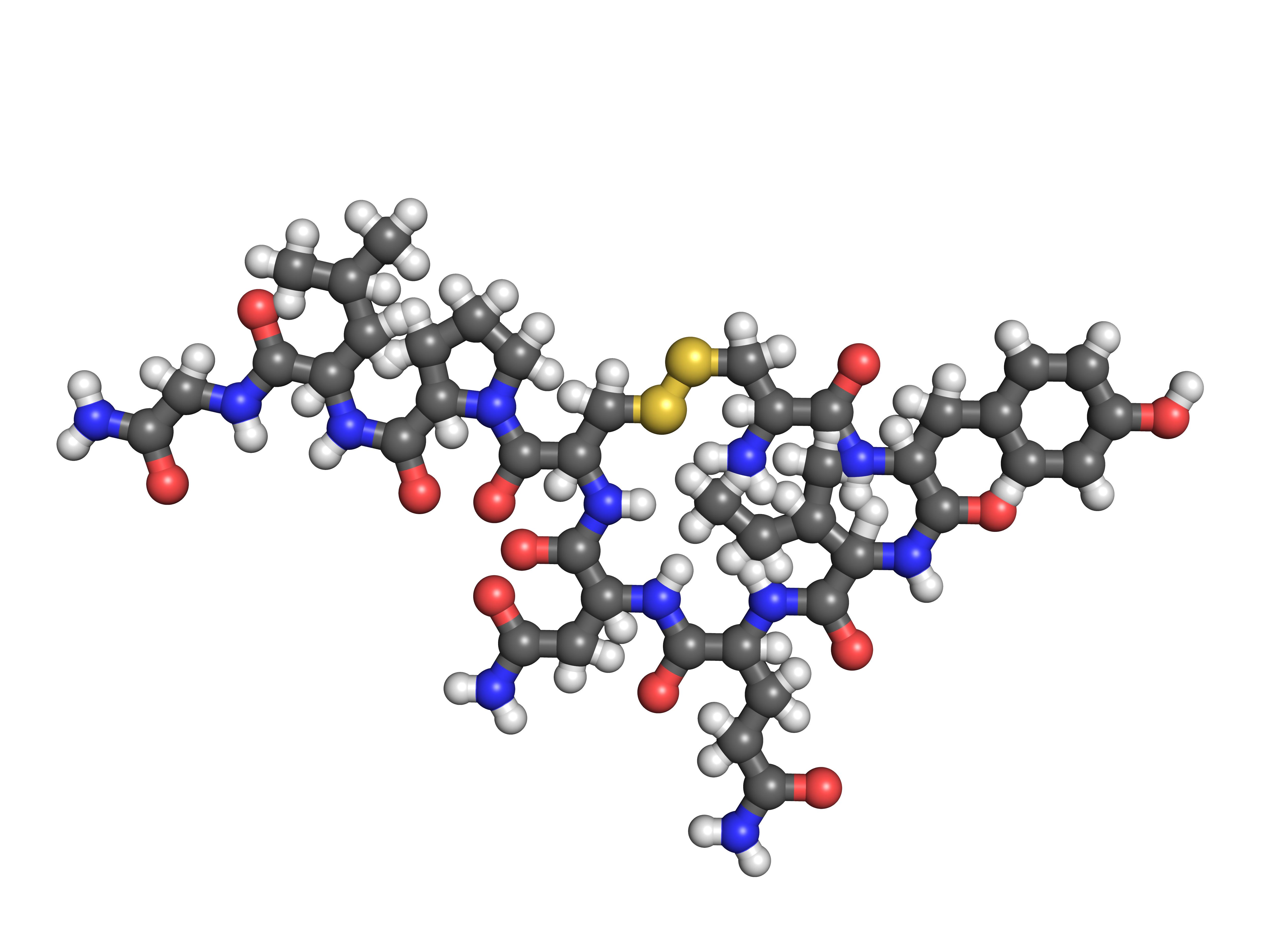
What is gamma-aminobutyric acid (GABA) in PTSD? GABA is the most important inhibitor of neurotransmitters in the central nervous system and is often dysfunctional in people with mood disorders. It has also been investigated in people with PTSD. GABA can be measured via peripheral levels in plasma, via central levels in cerebrospinal fluid, and in brain regions using proton magnetic resonance spectroscopy. What is the evidence for changes in GABA in people with PTSD? Moderate to low quality evidence found no significant differences in brain GABA levels between people with PTSD and controls. August 2021 Image: ©Molecular Science – stock.adobe.com

Hypothalamic-Pituitary-Adrenal axis
What is HPA dysfunction and PTSD? The biological response to stress is mediated through the Hypothalamic-Pituitary-Adrenal (HPA) axis and the sympathetic nervous system. Cortisol and the steroid hormone dehydroepiandrosterone (DHEA) and its sulfate form DHEA-S are important for elucidating the role of HPA dysfunctions in PTSD. Stress is a threat to the body’s ability to regulate internal processes following exposure to an adverse event. People adapt physiologically and behaviourally in response to stress to re-establish internal balance. Altered HPA axis activity can be detrimental to physical and psychological health. What is the evidence for HPA dysfunction in people with PTSD?…

Immune system
What is the immune system in PTSD? The immune system is the body’s first line of defence. It uses proteins called cytokines that are secreted by immune cells to allow cell-to-cell communication. Cytokines include interleukins (IL), interferons (IFN), tumor necrosis factors (TNF), transforming growth factors (TGF), and chemokines. These have influence over many cell types, including T helper lymphocytes (Th cells, or white blood cells). Cytokines act to regulate immunological and inflammatory responses to pathogens. They function as intermediaries between the immune system and the central nervous system. C-reactive protein (CRP) is released by the body during inflammation. Increased CRP…
Neurometabolites
What are neurometabolites in PTSD? Metabolites include N-acetylaspartate (NAA), creatine (Cr), trimethylamines/ choline containing compounds (Cho) and glutamine (Gln). These derivatives are indirect indicators of biochemical activity. Alteration in levels of NAA/Cr ratio is associated with reduction in the protective myelin sheath surrounding neurons. Decreased levels of NAA are associated with neuron death, or injury to the part of the neuron that connects to other cells, the axon. Alterations in some neurometabolites have been found in bipolar disorder and schizophrenia, and these have been investigated in PTSD. What is the evidence for neurometabolite alterations in people with PTSD? Moderate quality…
Neuropeptides
What are neuropeptides in PTSD? Neuropeptides are a class of molecules that can modulate the activity of neurotransmitters to increase or decrease the strength of synaptic signalling. Different neuropeptides are involved in different brain functions such as reward, food intake, metabolism, reproduction, social behaviours, and learning. Peripherally, they function like peptide hormones and modulate many bodily functions. What is the evidence for neuropeptides in people with PTSD? Moderate quality evidence found a large effect of lower neuropeptide Y levels in people with PTSD compared to controls without PTSD. This effect was also found in non-medicated samples. High quality evidence found…

Neurotrophins
What are neurotrophins in PTSD? Neurotrophins, such as brain-derived neurotrophic factor (BDNF) and nerve growth factor, are altered in bipolar disorder and schizophrenia. They regulate neuronal survival and growth during development. Effects of neurotrophins on neuronal transmission in the hippocampus, cortex, cerebellum, and basal forebrain are important for learning and memory processes. What is the evidence for changes in neurotrophins in people with PTSD? Moderate to low quality evidence found no significant differences in BDNF levels between people with PTSD and controls without PTSD. August 2021 Image: ©Giovanni Cancem – stock.adobe.com
Green - Topic summary is available.
Orange - Topic summary is being compiled.
Red - Topic summary has no current systematic review available.