Frontal lobe
What is the frontal lobe?
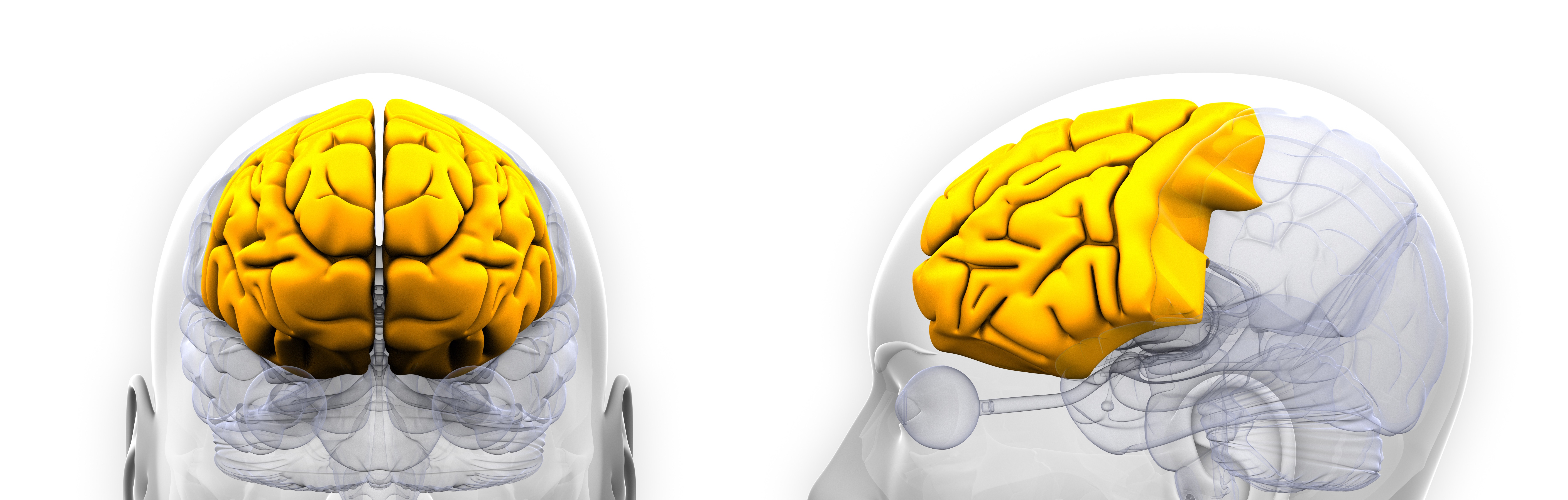
The frontal lobe comprises the anterior portion of the brain and is anatomically defined by four key gyri – the superior, middle, inferior and medial frontal gyri. The prefrontal cortex forms the rostral pole of the frontal lobe and is one of the most highly developed brain regions. The frontal lobe and its regions have widespread connections throughout the brain, particularly the prefrontal cortex. Proposed functions of the prefrontal cortex are involved mainly with executive functions and higher level cognition, such as working memory, problem solving and planning. The prefrontal cortex has also been implicated as a storage site for declarative memory such as semantic and episodic knowledge. This region has reciprocal connectivity with the amygdala, and is in a position to use experience and learning to influence behavioural responses and evaluate situations. The most posterior section of the frontal lobe is the pre-central gyrus, the primary motor cortex, also surrounded by associative and supplementary motor regions.
What is the evidence for frontal lobe alterations?
Structural changes
High quality evidence found schizophrenia is associated with significant reductions in grey and white matter volume of the frontal lobe, with greater reductions over time in people with schizophrenia than in controls. Specifically, moderate to high quality evidence found reduced grey matter in the prefrontal cortex, left orbito-frontal gyrus, left superior frontal gyrus, and bilateral medial, middle and inferior frontal gyri in chronic patients. There was also an absence of normal leftward asymmetry in the Sylvian fissure, and a higher frequency of abnormal (reversed) asymmetry in the frontal lobe of patients.
People with first-episode schizophrenia showed reduced grey matter in inferior, middle and medial frontal and precentral gyri. There was decreased right superior frontal grey matter in medication-naïve first-episode patients and increased right superior frontal grey matter in treated first-episode patients. A high risk of schizophrenia was particularly associated with reduced grey matter in superior and inferior frontal gyri.
Functional changes
Moderate quality evidence found increased activation during auditory hallucinations in the inferior and superior frontal gyri, and decreased activation during auditory tasks in the superior frontal gyrus of people with schizophrenia. Compared to controls without schizophrenia, there was decreased activation during cognitive control tasks in the right middle/inferior frontal gyrus and bilateral middle frontal gyrus. During timing tasks, there was increased activation in the right inferior frontal gyrus. During executive functioning tasks, there was decreased activation in the middle and medial frontal gyri, and decreased activation in the superior and inferior frontal gyri. During episodic memory encoding, there was reduced activity in the right superior frontal gyrus and bilateral inferior frontal gyri, and increased activity in the left precentral gyrus. During episodic memory retrieval, there was reduced activity in the left inferior frontal gyrus and left middle frontal gyrus, but increased activity in the left precentral gyrus and right middle frontal gyrus. During emotion processing tasks, there was reduced activity in the superior frontal gyrus. There was decreased activity in the inferior frontal gyrus and increased activity in the medial to superior prefrontal gyrus during explicit threat processing of facial stimuli. There was decreased activity in the medial prefrontal cortex and left orbito-frontal cortex during theory of mind tasks. Following cognitive remediation, patients showed increased activity in the left middle frontal gyrus, left inferior frontal gyrus, left superior frontal gyrus, and medial frontal gyrus. There was decreased activation in the left middle frontal gyrus during reward anticipation tasks and decreased activation in the right inferior frontal gyrus during empathy tasks.
In first-degree relatives of people with schizophrenia, there was decreased resting-state brain activity in the right inferior frontal gyrus compared to controls. The right inferior frontal gyrus showed increased activation during cognitive tasks and decreased activation during emotion tasks in relatives. There was also increased activation in the right superior frontal gyrus and decreased activation in the left medial frontal gyrus during emotion tasks. There was decreased activity in the right middle frontal gyrus and right inferior frontal gyrus, and increased activity in the right frontopolar region during working memory tasks in relatives.
Moderate quality evidence found decreased phosphomonoester (PME) levels in the prefrontal cortex of people with first-episode psychosis and people with schizophrenia when compared to controls. There were increased phosphodiester (PDE) levels in the prefrontal cortex of first-episode patients. Moderate to low quality evidence found decreased PME and increased PDE levels in the frontal lobe of first-degree relatives of people with schizophrenia.
Moderate quality evidence found N-acetylaspartate (NAA) and creatine (Cr) levels were reduced in frontal grey and white matter, particularly the prefrontal cortex and frontal pole, in both first episode and chronic schizophrenia. NAA/Cr ratio was reduced in the prefrontal cortex of people at clinical or familial risk of schizophrenia.
Moderate to high quality evidence found reduced glutamate (Glu) and increased glutamine (Gln) levels in the frontal cortex of people with schizophrenia. There was a medium-sized increase in Glu+Gln in the medial prefrontal cortex of unmedicated people with schizophrenia, and increased Glu/Gln ratio and glutamate+glutamine levels in the frontal lobe of first-degree relatives of people with schizophrenia.
High quality evidence found a small decrease in myo-inositol levels in the medial prefrontal region in people with schizophrenia, while moderate quality evidence found reduced translocator protein.
October 2020
Fact Sheet Technical Commentary
Green - Topic summary is available.
Orange - Topic summary is being compiled.
Red - Topic summary has no current systematic review available.