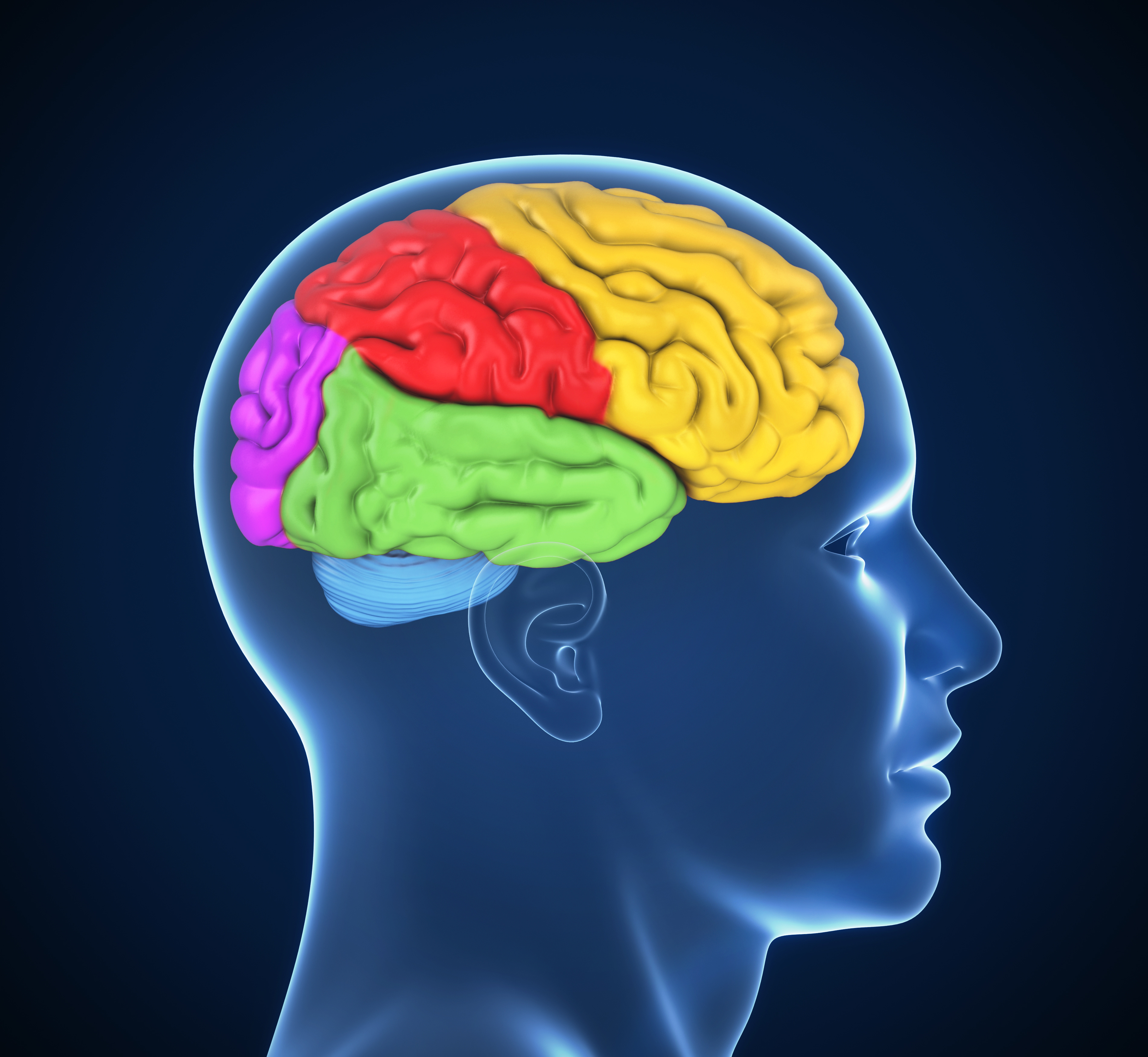
Brain regions
This category summarises the information contained in the brain structure and function sections of the library, presented according to specific regions of the brain. Most regions show some alteration, with much research investigating the frontal, temporal, hippocampal, and amygdala regions, as well as whole brain volume. Click on the links and tabs below to access the information, or browse via the drop-down menu on the left.
Image: ©koya979 – stock.adobe.com
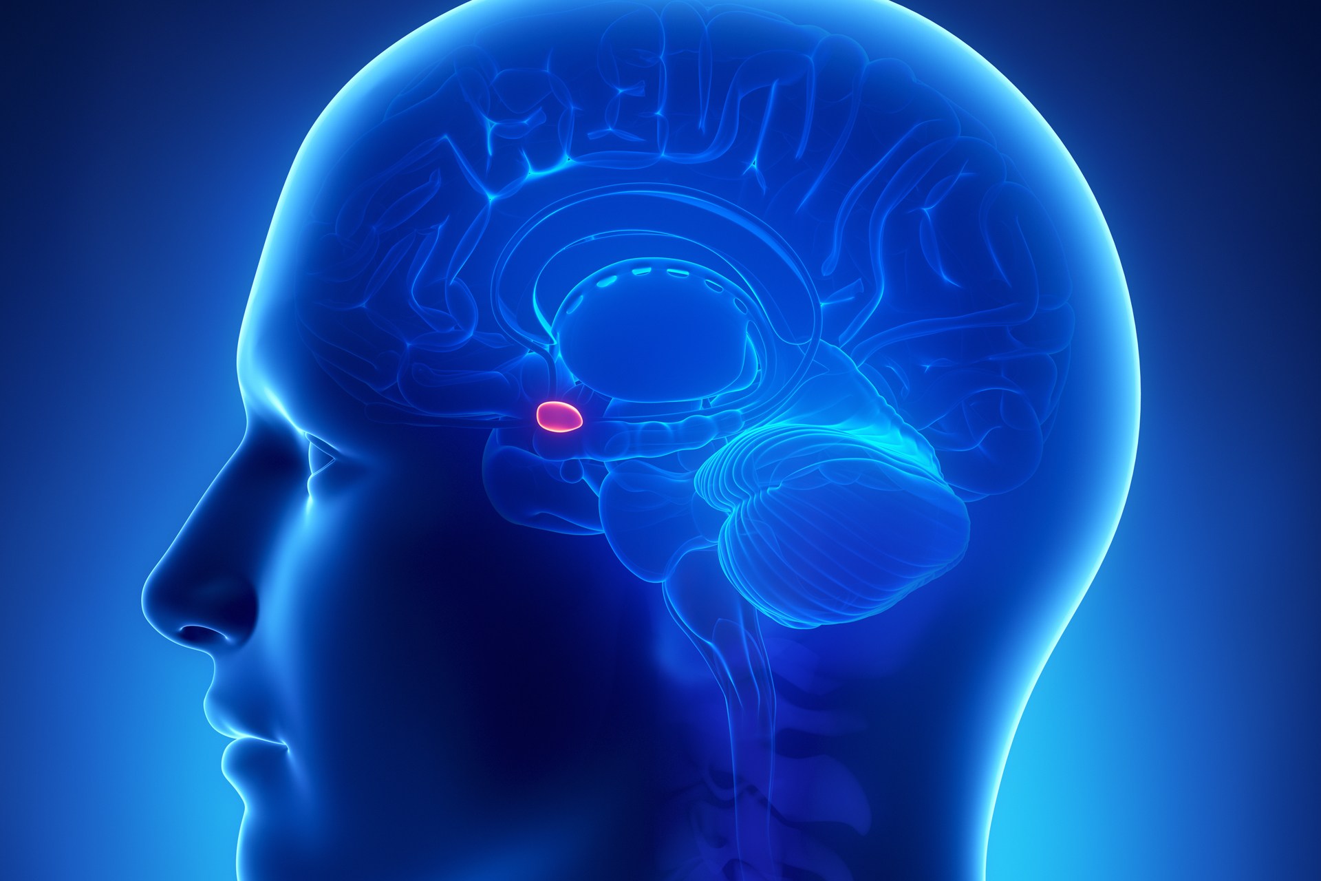
Amygdala
What is the amygdala? The amygdala is located deep in the medial temporal lobe, and has reciprocal connections with many regions of the cortex, such as prefrontal and cingulate cortex, as well as sub-cortical regions such as the brainstem and hippocampus. The amygdala is implicated in the processing and memory of emotional responses, particularly emotional learning, as well as mediating the autonomic expression of emotion. What is the evidence for amygdala alterations? Moderate to high quality evidence found reduced grey matter volume in the amygdala and the amygdala-hippocampus region of people with schizophrenia compared to controls. There were also reductions…

Arcuate fasciculus
What is the arculate fasciculus? The arcuate fasciculus is a bundle of axons that connects the temporal cortex and inferior parietal cortex to locations in the frontal lobe. One of the key roles of the arcuate fasciculus is connecting Broca’s and Wernicke’s areas, which are involved in producing and understanding language. Therefore, there may be anomalies in the arculate fasciculus of people with schizophrenia who experience auditory-verbal hallucinations. What is the evidence for changes in the arculate fasciculus? Moderate quality evidence found reduced white matter integrity in the bilateral arcuate fasciculus, including bilateral anterior and posterior segments, and the left…
Basal ganglia
What is the basal ganglia? The basal ganglia is a group of sub-cortical nuclei thought to be involved in motor control and learning. The nuclei comprising the basal ganglia include the caudate, putamen, globus pallidus, the subthalamic nucleus, and the substantia nigra. The caudate and putamen together form the striatum, while the globus pallidus and the putamen together form the lenticular nucleus. The striatum is the principal input centre, receiving afferents primarily from the cortex, but also from the substantia nigra, thalamus, and external globus pallidus. There are two primary pathways from the striatum through the basal ganglia (‘direct’ and…
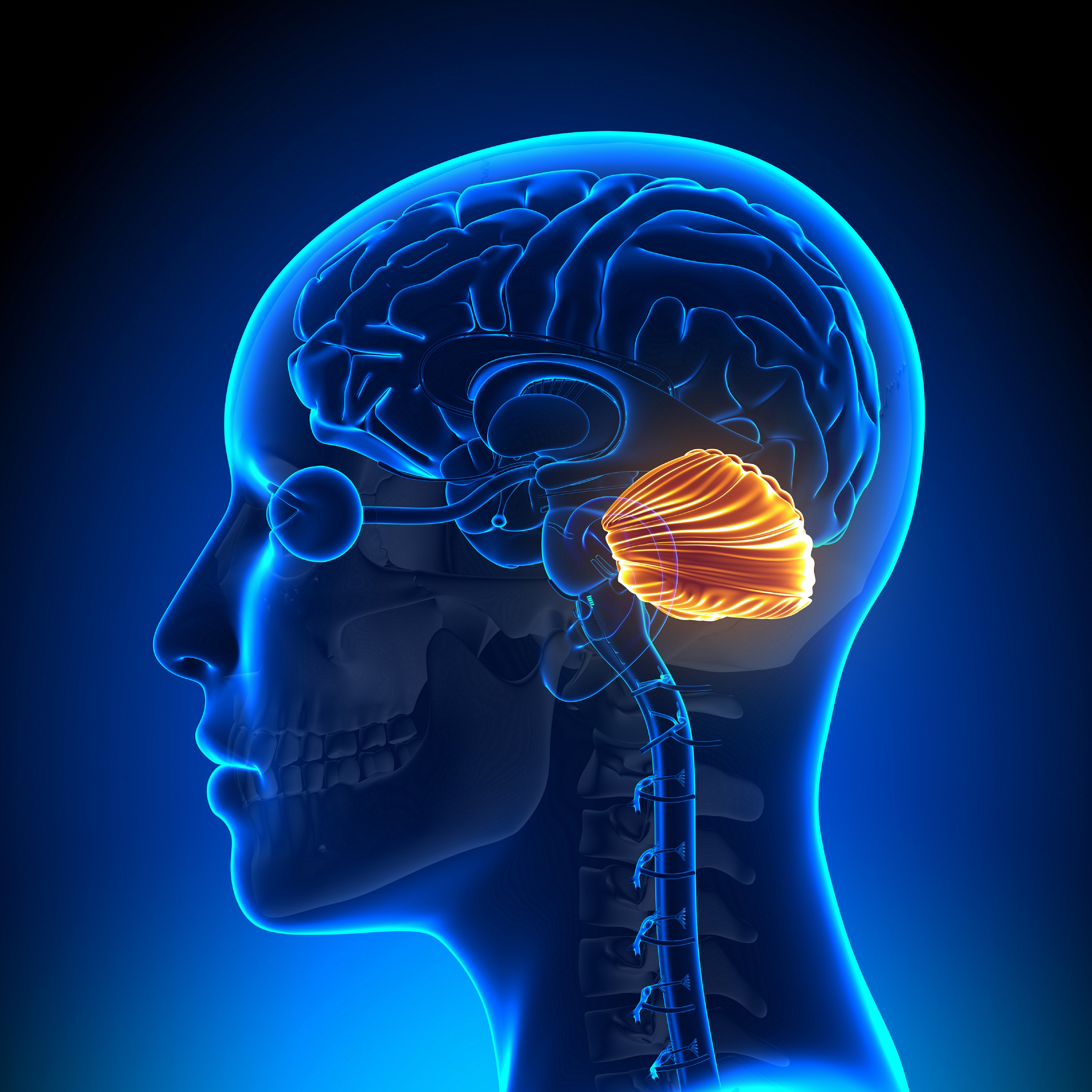
Cerebellum
What is the cerebellum? The cerebellum sits below the larger cerebrum of the brain, and is connected via the brainstem. The cerebellum is divided into two hemispheres separated dorsally by a midline zone called the vermis. It contains three primary lobes, the flocculonodular lobe, anterior lobe, and posterior lobe. Broadly, the cerebellum is thought to function in fine motor control (coordination and precision) and motor learning, balance, posture, as well as some cognitive and emotional capacity. The interaction of sensory, cognitive and motor functions may also contribute to proprioception (the awareness of self in space), planning movements, and evaluating information…
Cingulate cortex
What is the cingulate cortex? The cingulate cortex is part of the medial frontal cortex, located immediately dorsal to the corpus callosum along the sagittal midline. The anterior cingulate cortex has three key divisions which may be functionally distinct (dorsal, rostral, subcallosal). The dorsal part of the anterior cingulate cortex has reciprocal connections with the prefrontal and parietal cortices as well as the frontal eye fields, and plays a primary role in balancing top-down and bottom-up processing of external stimuli; that is, monitoring behaviour and incoming stimuli in the context of current goals, and assigning control to other areas in…
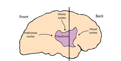
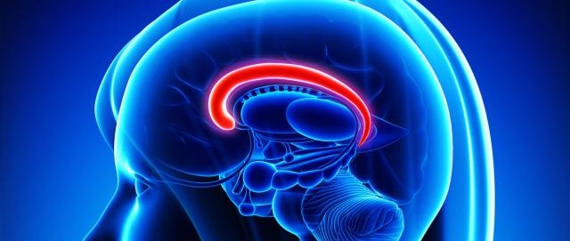
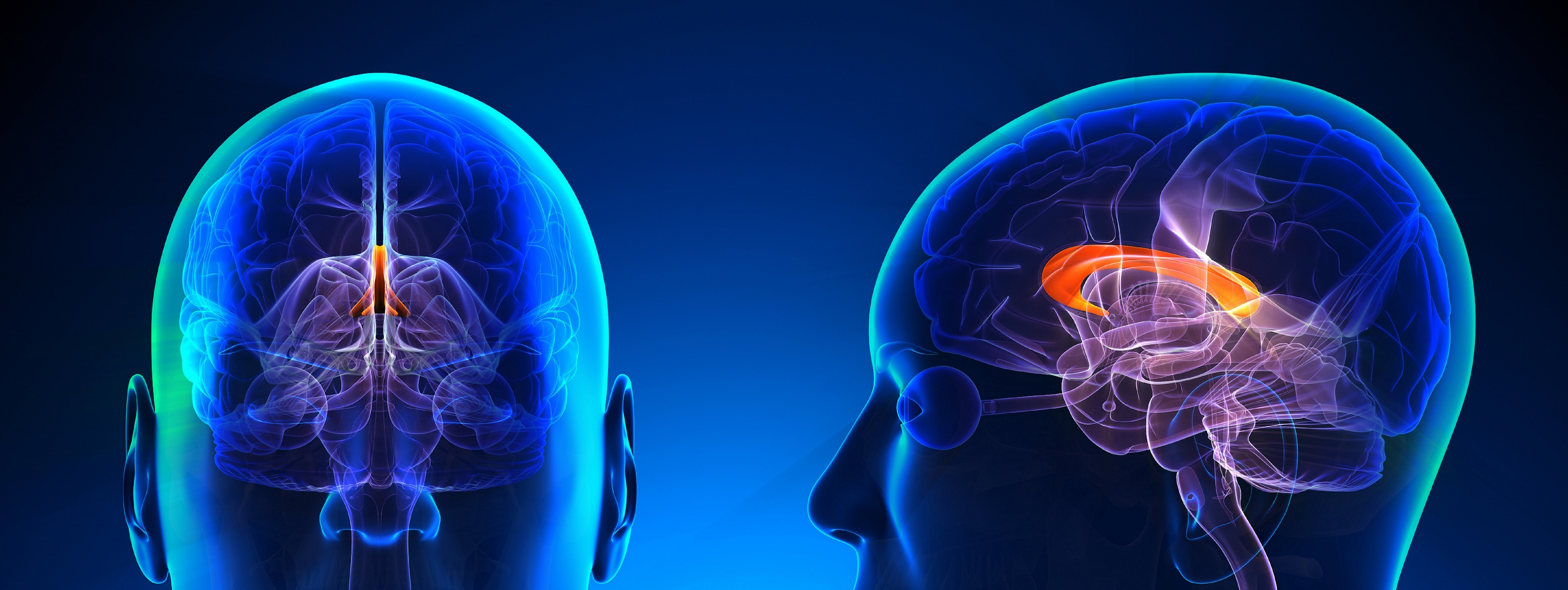
Claustrum
What is the claustrum? The claustrum is a thin irregular sheet of grey matter located sagitally between the external capsule and the extreme capsule fibre tracts. The connectivity of the claustrum has not been extensively determined, however it appears to have connections with almost all cortical regions, as well as some subcortical connections such as the hippocampus, amygdala and basal ganglia. The function of the claustrum is also largely unclear, but may be involved in some functions of the neighbouring insula. The widespread connectivity of the claustrum places it in a prime position for multimodal integration and processing of perceptual,…
Corpus callosum
What is the corpus callosum? The corpus callosum is the bundle of inter-hemispheric white matter tracts that plays an essential role in the transfer and integration of sensory, motor and cognitive information between homologous regions in opposite hemispheres. It is the primary source of contralateral connections between the hemispheres and contains as many as 250 million axons. What is the evidence for corpus callosum alterations? Moderate to high quality evidence found volume and white matter reductions in the corpus callosum of people with schizophrenia compared to controls. There were reductions in frontal white matter via genu of the corpus callosum…
Default mode network
What are default mode network dynamics? The ‘default mode’ system refers to a network of regions including the precuneus, posterior cingulate cortex, medial prefrontal cortex, and medial, lateral and inferior parietal cortices, that appear to be active in the resting brain, and consistently show attenuations of activity following onset of a task-related activity. Default mode network (DMN) attenuation is not task specific; however the magnitude of reduction is dependent on the cognitive load and task requirements. The more demanding the task being performed, the stronger the deactivation. DMN activity is characterised by coherent low frequency (less than 0.1 Hz) neural…
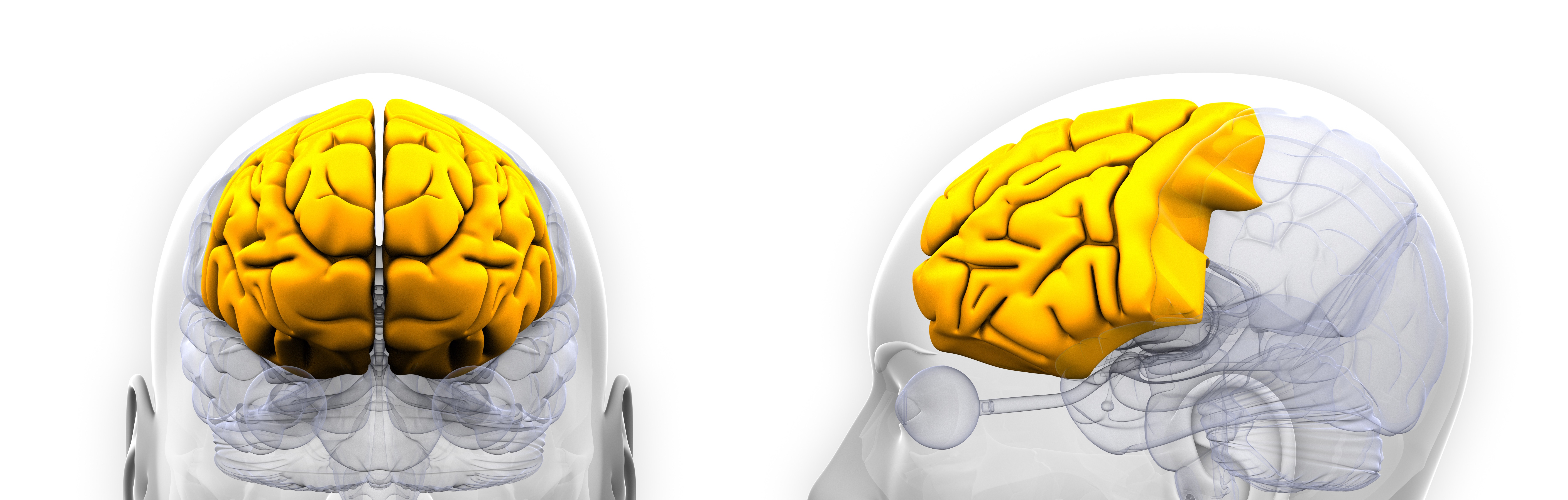
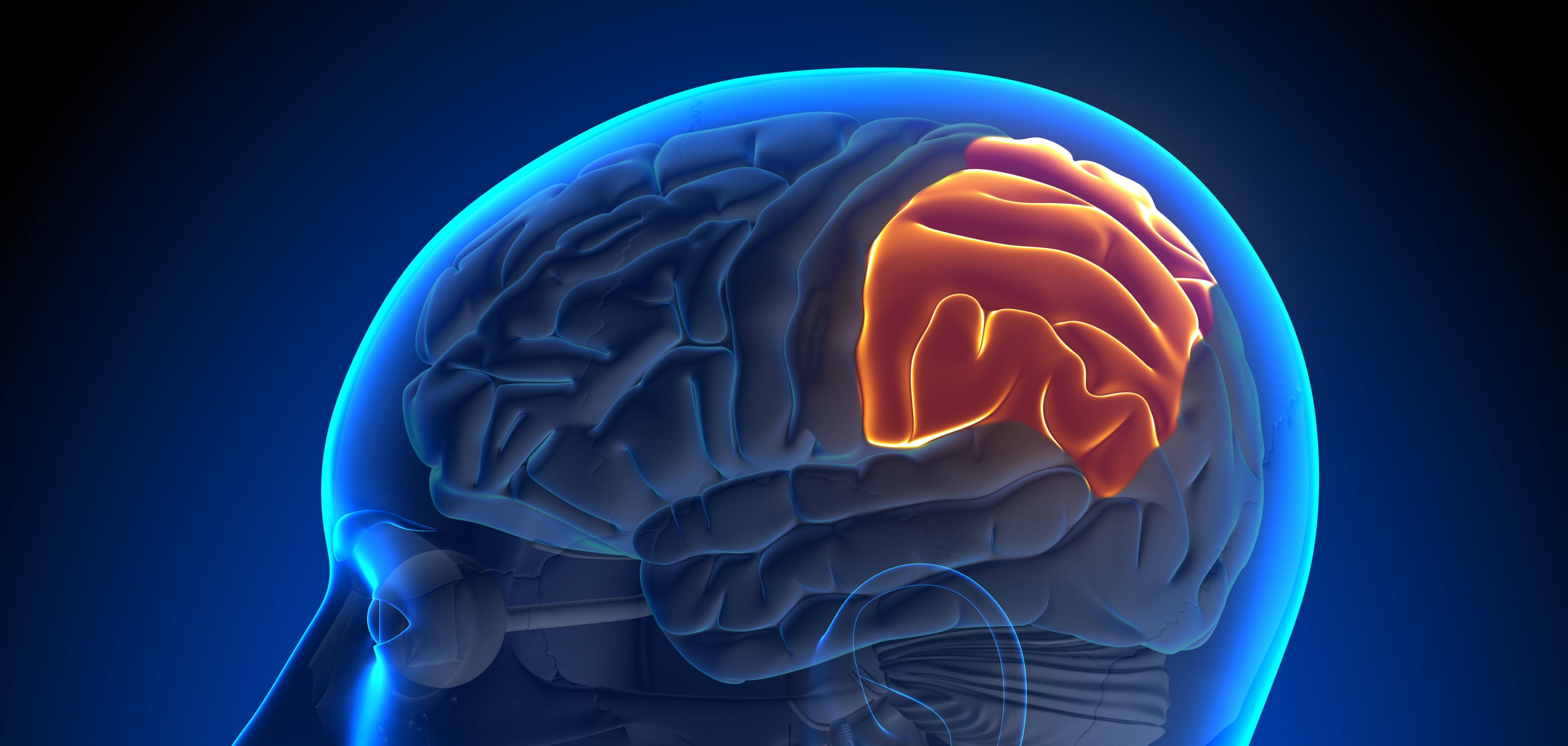
Frontal lobe
What is the frontal lobe? The frontal lobe comprises the anterior portion of the brain and is anatomically defined by four key gyri – the superior, middle, inferior and medial frontal gyri. The prefrontal cortex forms the rostral pole of the frontal lobe and is one of the most highly developed brain regions. The frontal lobe and its regions have widespread connections throughout the brain, particularly the prefrontal cortex. Proposed functions of the prefrontal cortex are involved mainly with executive functions and higher level cognition, such as working memory, problem solving and planning. The prefrontal cortex has also been implicated…
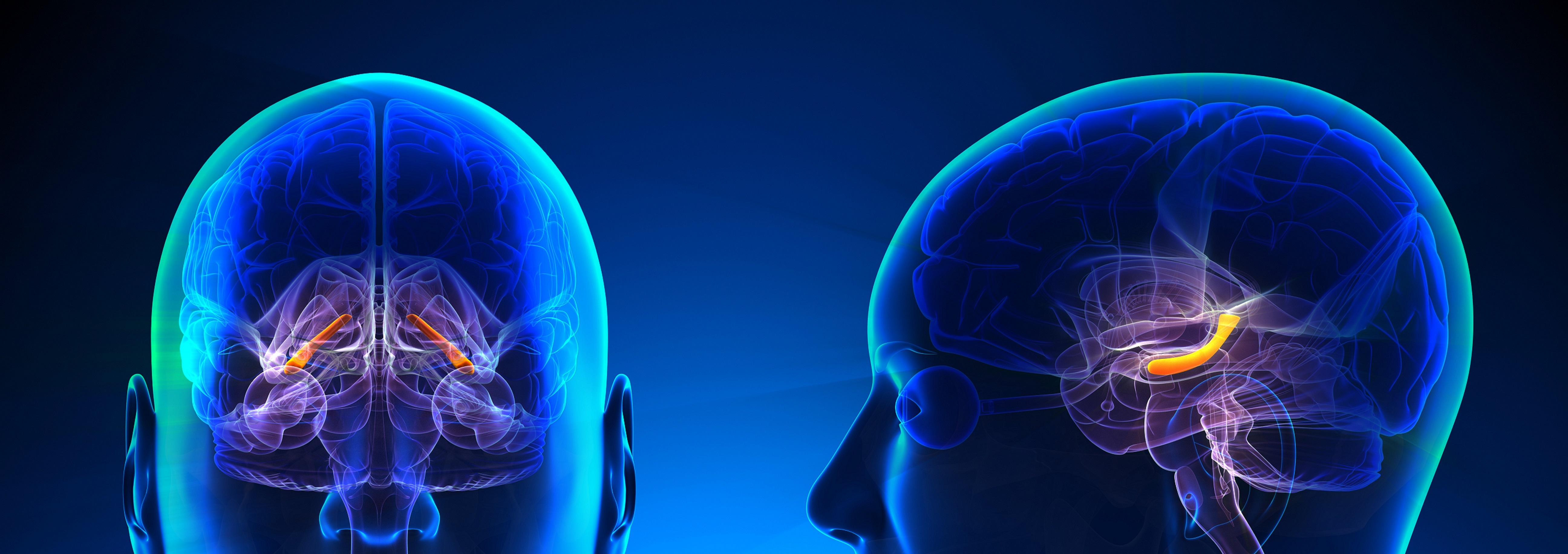
Hippocampus
What is the hippocampus? The hippocampus is located deep within the medial temporal lobe and has extensive connections, largely to cortical association areas including the sensory modalities. This widespread connectivity facilitates multimodal integration of sensory information, and likely contributes to the role of the hippocampus in generating memory and facilitating spatial navigation. The medial temporal lobes, particularly the hippocampus and the surrounding cortical regions, have been implicated as crucial facilitators in the formation of new declarative memories. What is the evidence for changes in the hippocampus? Structural changes Moderate or high quality evidence found hippocampal and parahippocampal grey and white…
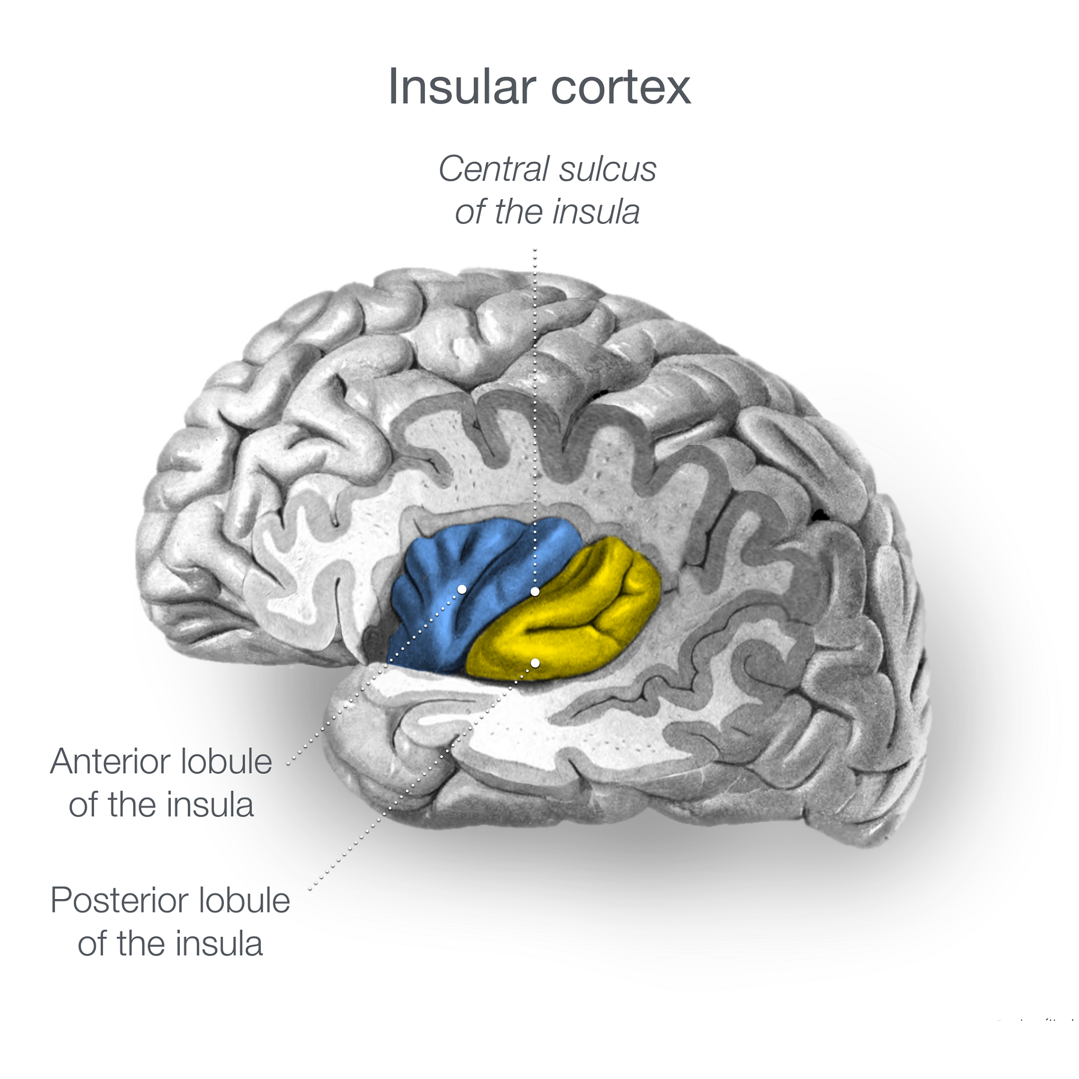
Insular
What is the insular? The insular cortex is located deep within the lateral fissure, between the frontal and temporal lobes. The insular has connections with the thalamus, amygdala and cortex, with suggested functions including the integration of sensory, affective and cognitive components of a painful stimulus to create the sensation of pain; homeostatic regulation; as well as motor control such as speech articulation; it has also been linked with internal awareness. What is the evidence for insular alterations? Structural changes Moderate to high quality evidence found reductions in insular grey matter volume in both chronic and first-episode schizophrenia (treated or…
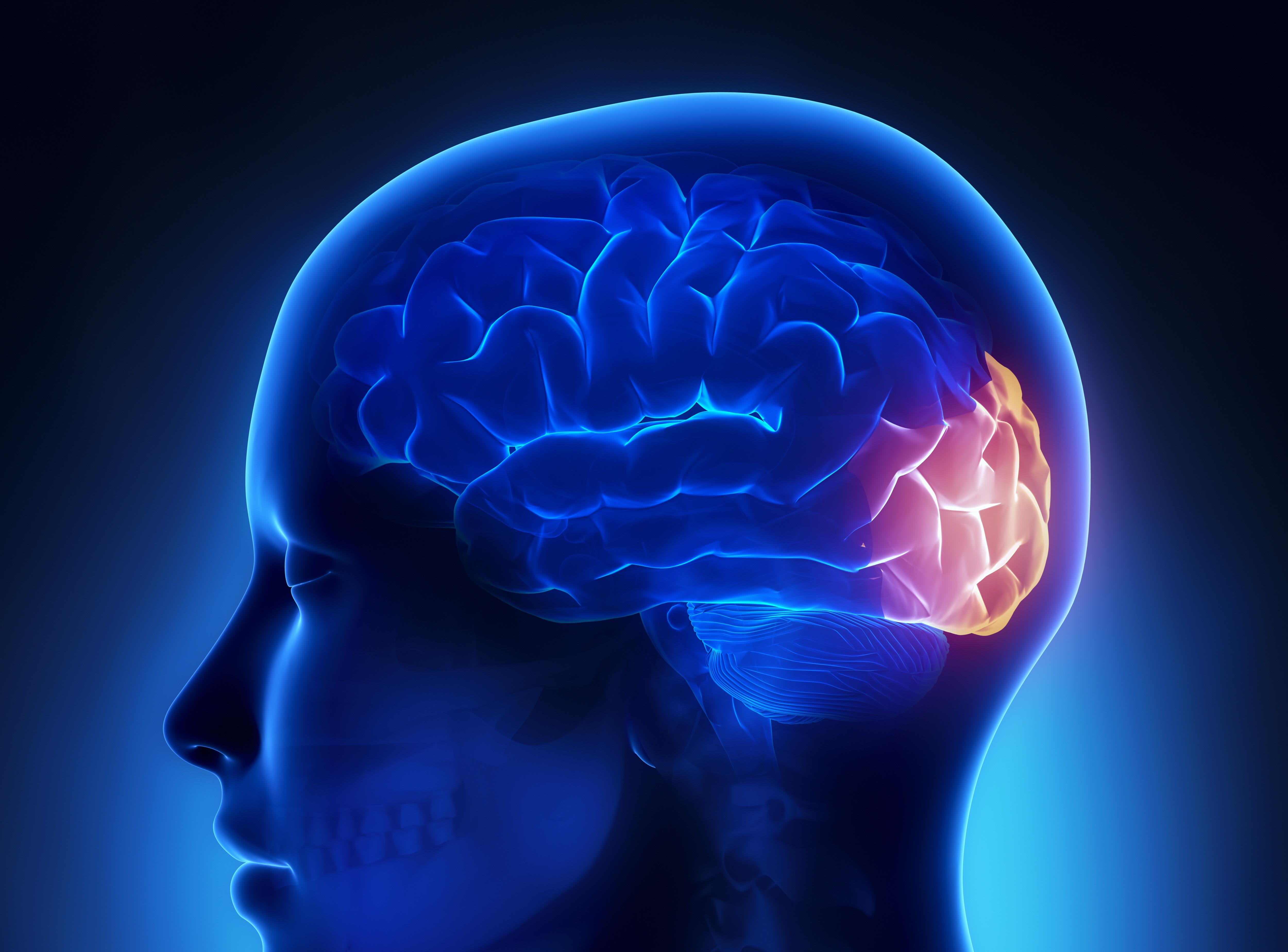
Occipital lobe
What is the occipital lobe? The occipital lobe is located at the posterior section of the brain and primarily comprises the brain’s visual cortices. There are two streams of visual information through the visual primary and association cortices, which deal separately with broad object details and motion, and fine detail and colours. What is the evidence for occipital lobe alterations? Structural changes Moderate to high quality evidence found reductions in occipital grey matter in people with schizophrenia compared to controls. Increased antipsychotic dose was associated with decreased occipital grey matter over time, and decreases were associated with lower overall functioning….
Parietal lobe
What is the parietal lobe? The parietal cortex is located posterior to the frontal lobe. It is structurally divided into the superior, middle and inferior gyri. The most anterior portion of the parietal lobe forms the post-central gyrus, the somatosensory cortex. Posterior to this are the parietal association regions and the visual regions of the posterior parietal cortex involved in visuospatial processing. What is the evidence for parietal lobe alterations? Structural changes Moderate to high quality evidence found decreased parietal grey matter volume in medicated people with schizophrenia compared to controls. There were reductions in the left inferior parietal gyrus…
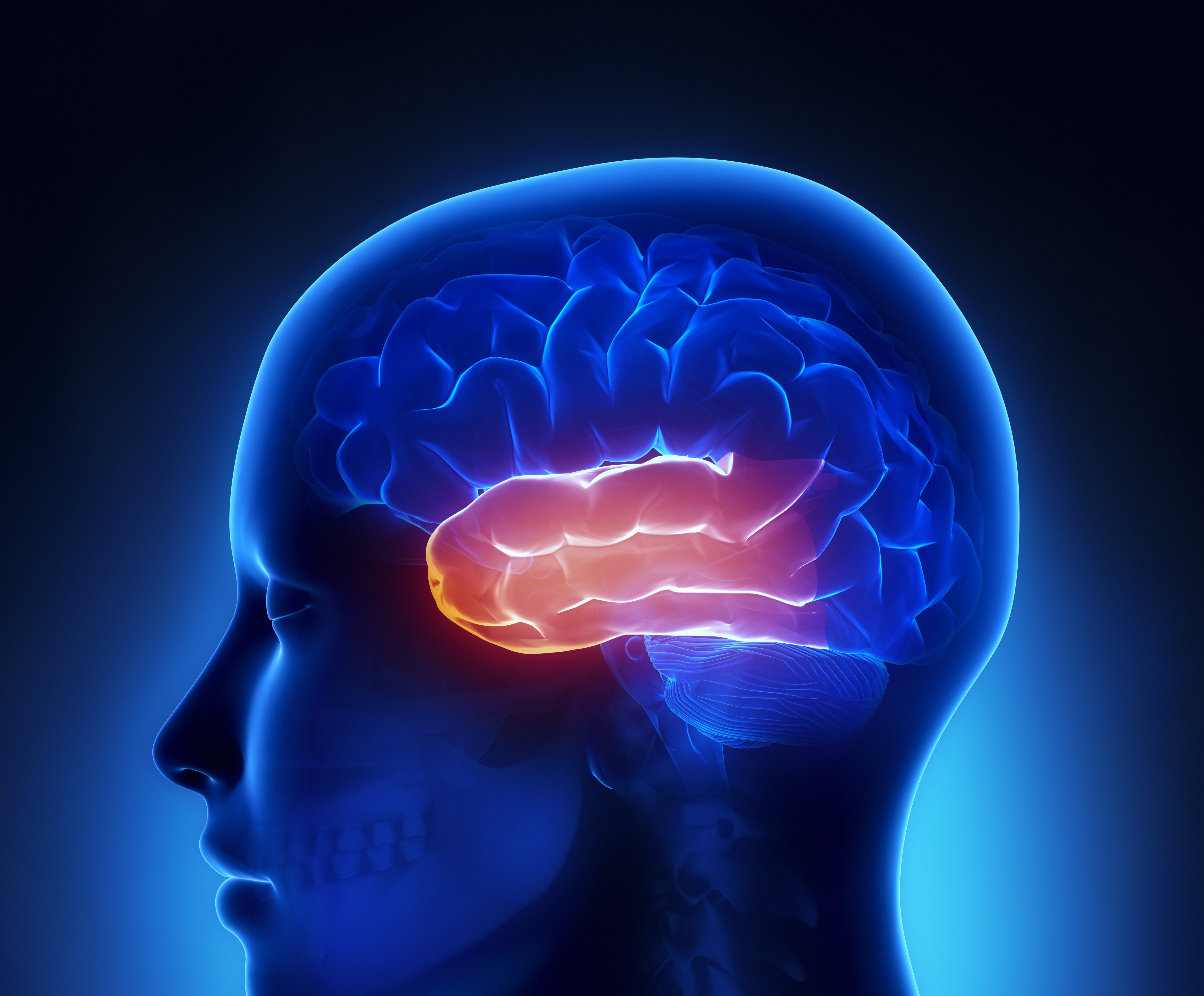
Temporal lobe
What is the temporal lobe? The temporal lobe is structurally divided into the superior, middle, inferior and medial gyri. The superior temporal gyrus comprises the primary auditory cortex, while nearby temporal regions function in higher level auditory processing, including speech and language. Inferior temporal regions are involved in higher level visual processing and the temporal-occipital gyrus is involved in face processing. The medial temporal lobe comprises the hippocampus and is thought be involved in the formation and propagation of memory. What is the evidence for temporal lobe alterations? Structural changes Moderate to high quality evidence found reduced grey and white…
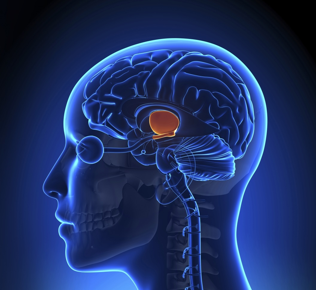
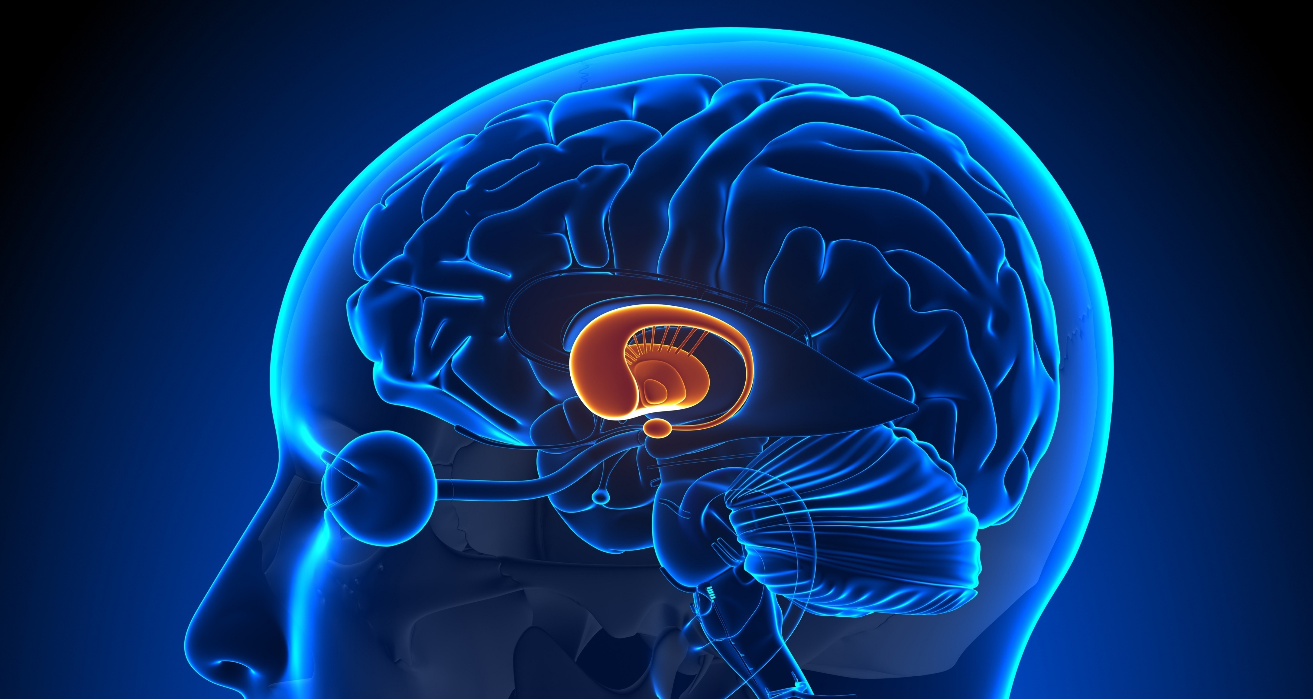
Thalamus
What is the thalamus? The thalamus is a midline structure located directly on top of the brainstem, surrounding the third ventricle. It is thought to be primarily involved in relaying information from the brainstem and subcortex into the cortex, particularly sensation, special sense and motor signals. Every sensory system except olfaction utilises a thalamic relay to the associated cortical region. The thalamus has also been implicated in autonomic functions such as the regulation of consciousness, sleep and wakefulness. The thalamus may also have higher cognitive functions, being implicated in some emotional processing as well as memory propagation. What is the…
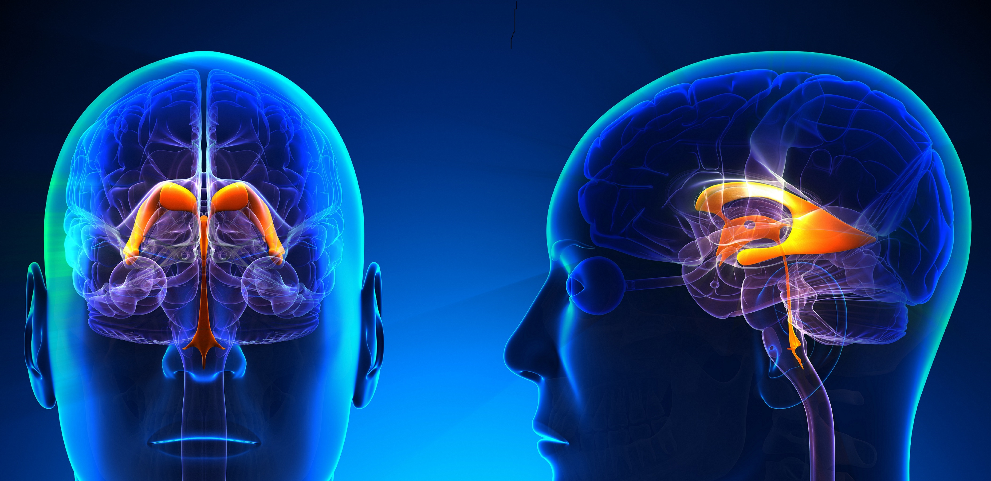
Ventricular system
What are the ventricles? The ventricular system of the brain functions to provide support to surrounding tissues with cerebrospinal fluid (CSF), produced in the choroid plexus tissue lining many of the ventricles. The system comprises the bilateral cerebral lateral ventricles, the midline third and fourth ventricles, and the central canal of the spinal cord. The lateral ventricles have four sections, the frontal (anterior) horns; temporal (inferior) horns; body; and occipital (posterior) horns. The interventricular foramen connects the lateral ventricles to the third ventricle, and the cerebral aqueduct connects the third ventricle to the fourth. The fourth ventricle is continuous with…
Whole brain volume
What is whole brain volume and functioning? Investigation of whole brain anomalies considers the collective volume of the entire brain in structural imaging, without considering regionally specific differences in the volume of any individual structures. Alternatively, whole brain imaging can also consider overall grey matter or white matter volume. Whole brain functioning assesses the degree of connectivity across multiple brain regions. What is the evidence for whole brain volume and functioning? Structural changes Moderate quality evidence found lower brain weight in people with schizophrenia compared to controls. Moderate to low quality evidence found male patients who died by suicide had…
Green - Topic summary is available.
Orange - Topic summary is being compiled.
Red - Topic summary has no current systematic review available.