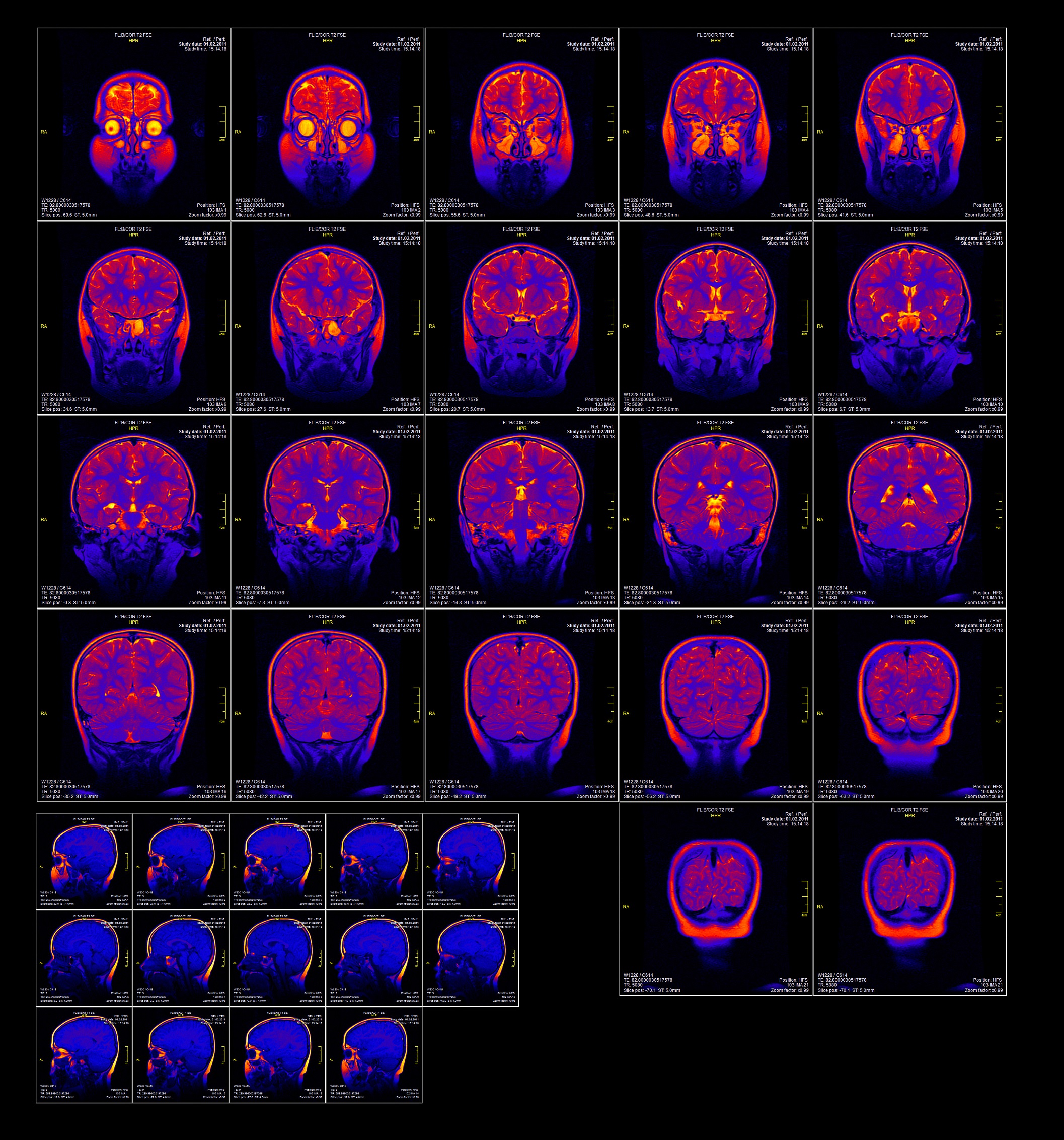
Functional magnetic resonance imaging
What is fMRI?
Functional magnetic resonance imaging (fMRI) is used to determine functional activation of brain regions when an individual performs tasks (or rests) inside an MRI scanner. Most commonly fMRI studies use visual, auditory, motor or sensory stimuli to evoke neural responses in the brain. Recent fMRI studies also examine activity of the brain at rest. Changes in blood flow are interpreted to represent brain activation (or deactivation) associated with a particular brain state (i.e, while performing a particular activity, or while the brain is at rest). Functional activity has been investigated in people with schizophrenia compared to people without schizophrenia to identify regions of increased or decreased brain function on the basis of blood flow.
What is the evidence for fMRI brain function?
Moderate quality evidence suggests decreased local organisation and small-worldness (balance of local organisation and global integration) in people with schizophrenia compared to controls. There was reduced connectivity within the default network (self-related thought), the affective network (emotion processing), the ventral attention network (processing of salience), the thalamus network (gating information) and the somatosensory network (sensory and auditory perception). There was reduced connectivity between the ventral attention network and the thalamus network, the ventral attention network and the default network, the ventral attention network and the frontoparietal network (external goal-directed regulation), the frontoparietal network and the thalamus network, and the frontoparietal network and the default network. There was increased connectivity between the affective network and the ventral attention network.
During executive functioning and working memory tasks, there was decreased activation in the frontal lobe, including the dorsolateral prefrontal cortex, and in neocortical regions, including the parietal and occipital cortices and bilateral claustrum, fusiform gyrus, and cerebellum, and in subcortical regions, including the right putamen, hippocampus and left mediodorsal thalamus. Moderate to low quality evidence suggests significant increases in functional activation in the anterior cingulate cortex, temporal lobe, parietal cortex, lingual gyri, insula and the amygdala.
During cognitive control tasks, there was decreased activation in the bilateral anterior cingulate/paracingulate gyrus, left inferior parietal gyrus, right middle/inferior frontal gyrus, bilateral middle frontal gyrus, right thalamus, and left cerebellum. There was increased activation in the right middle occipital and bilateral precentral gyri.
During timing tasks, there was decreased activation in the bilateral caudate nuclei, left middle occipital gyrus, right inferior occipital gyrus, bilateral supplementary motor area, and right putamen. There was increased activation during timing tasks in bilateral superior parietal gyri, right inferior frontal gyrus, and right middle temporal gyrus.
During memory encoding tasks, there was decreased activation in the medial frontal gyri and the hippocampus. During memory retrieval tasks, decreased activation was seen in the medial and inferior frontal gyri, the cerebellum, hippocampus, and the fusiform gyrus, with increases in the anterior cingulate cortex and the medial temporal gyrus. During episodic memory encoding, there was decreased activation in the right superior frontal gyrus, bilateral inferior frontal gyri, right inferior parietal gyrus, right lingual gyrus, left hippocampus, and right posterior cingulate. There was increased activation in the left precentral gyrus, left middle temporal gyrus, left post-central gyrus, left cingulate and left parahippocampal gyrus. During episodic memory retrieval, there was decreased activation in the left inferior frontal gyrus, left middle frontal gyrus, right cuneus, right cingulate gyrus, bilateral thalamus, and bilateral cerebellum. There was increased activaton in the left precentral gyrus, right middle frontal gyrus, right thalamus and right parahippocampal gyrus.
During emotion processing tasks, there was decreased activation in the parahippocampus, superior frontal gyrus, middle occipital gyrus, fusiform gyrus, lentiform nucleus, and thalamus. There was increased activation in left amygdala, left hippocampus, left medial frontal region, left cuneus, and bilateral parietal cortex. During explicit threat processing, there was decreased activation in the inferior frontal gyrus, right cerebellum lobule VI, left fusiform gyrus, and thalamus, and increased activation in the medial prefrontal gyrus to superior prefrontal gyrus. During implicit threat processing, there was decreased activation in bilateral amygdala extending into putamen, hippocampus and parahippocampal gyrus, and fusiform gyrus extending into the cerebellum lobule IV/VI.
During theory of mind tasks, there was decreased activation in the medial prefrontal cortex (frontal medial and paracingulate), right premotor cortex (central opercular, postcentral, precentral), medial occipitoparietal, right lingual gyrus, left orbitofrontal cortex, left lateral occipitotemporal, left cingulate gyrus, and left middle temporal gyrus. There was increased activation in the left inferior parietal cortex and right inferior parietal cortex. During empathy tasks, there was decreased activation the right inferior frontal gyrus.
During inhibition tasks, there was decreased activation in the anterior and middle cingulate cortex, and increased activation in parietal and occipital regions. There was also decreased activation in the basal ganglia and inferior frontal cortex, and increased activation in the superior temporal gyrus during inhibition tasks.
During attention tasks, there was decreased activation in the anterior and middle cingulate cortex and the basal ganglia, and increased activation in the left supramarginal gyrus.
During linguistic tasks (mostly semantic reading), there was decreased activation in the lateral temporal regions and left putamen, and increased activation in bilateral frontal cortex and left putamen.
During reward stimuli tasks, there was decreased activation in the right ventral striatum.
During auditory hallucinations, there was increased activation in Broca’s area of the temporal lobe, insula, hippocampus, left parietal operculum, left and right postcentral gyrus, and left inferior frontal gyrus, and decreased activation of Broca’s area, the left middle temporal gyrus, left premotor cortex, anterior cingulate cortex, and left superior temporal gyrus.
In people with schizophrenia and formal thought disorder, moderate quality evidence found functional alterations (hyperactivation or hypoactivation) in the left superior and middle temporal gyrus.
Following cognitive remediation (40 session over 10 weeks), moderate to low quality evidence found increased activation in the left middle frontal gyrus, left inferior frontal gyrus, left superior frontal gyrus, pre- and postcentral gyrus, bilateral insula, parietal lobe, and medial frontal gyrus.
October 2020
Fact Sheet Technical Commentary
Green - Topic summary is available.
Orange - Topic summary is being compiled.
Red - Topic summary has no current systematic review available.
