
Brain structure
Brain structural changes have been found in people with schizophrenia. These alterations are have been found using magnetic resonance imaging (MRI), diffusion tensor imaging (DTI), optical coherence tomography (OCT), and computed tomography. Neuronal changes, and changes in brain weight, have also been observed. Click on the links or the tabs below to access the information, or browse the drop-down menu to the left.
Image: ©vegefox.com – stock.adobe.com

Brain weight
What is brain weight? Brain weight refers to the basic mass measurement of a post-mortem brain, either at time of autopsy (‘fresh’) or after formalin fixation (‘fixed’). Its ease of collection means it is a routine measurement at autopsy and has become a widely used tool for insight into brain integrity. If brain weight is altered, this provides a non-specific indication of neuropathology. It is often a presumed equivalent of the MRI findings of decreased brain volume. What is the evidence for brain weight? Moderate quality evidence suggests the brain of a person with schizophrenia is significantly lower in weight…
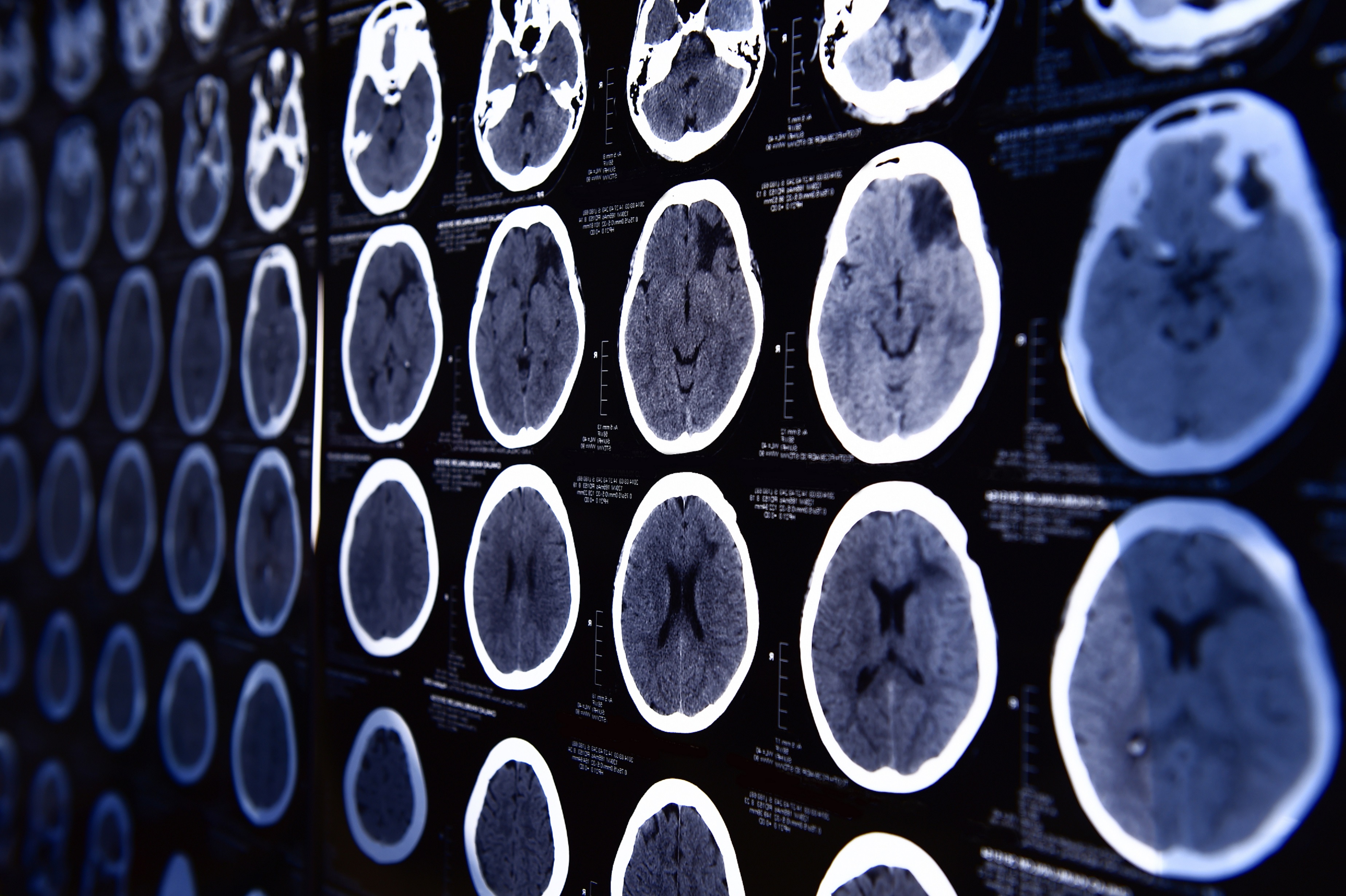
Computed tomography
What is computed tomography (CT)? CT imaging is a method for visualising the structural organisation of the brain using the attenuation of X-rays to generate image contrast. Tissues in regions of interest are highlighted based on their X-ray absorption properties, as dense tissues attenuate X-rays more than soft tissues, and air attenuates the least. Three-dimensional images are generated from a series of two-dimension X-ray images taken around a single axis of rotation. What is the evidence for CT brain structure? Moderate quality evidence suggests reduced temporal lobe volume in people with schizophrenia. Moderate to low quality evidence is unclear as…
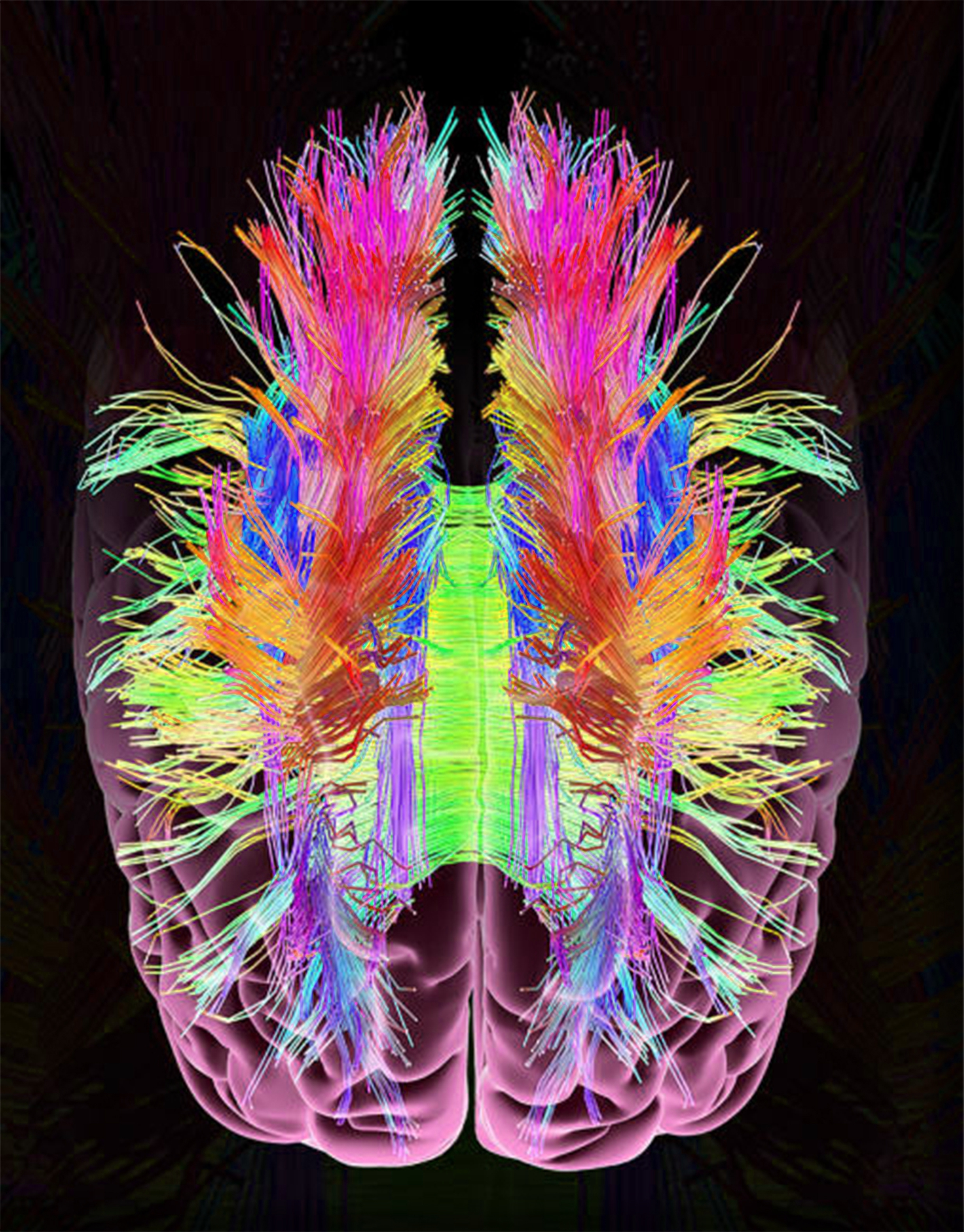
Diffusion tensor imaging
What is diffusion tensor imaging (DTI)? DTI is a specialised imaging technique that uses MRI technology to investigate the movement of water within tissues of interest. By applying a magnetic field, the movement (“diffusivity”) of water molecules can be visualised in vivo. The diffusion of water is influenced by the cellular structure of the surrounding tissues, and measures such as fractional anisotropy (FA) were derived as an approximate measurement for the freedom of movement. In areas of high structural coherence such as white matter, FA is highest, indicating that water is moving in relatively fixed directions. It is lower in…
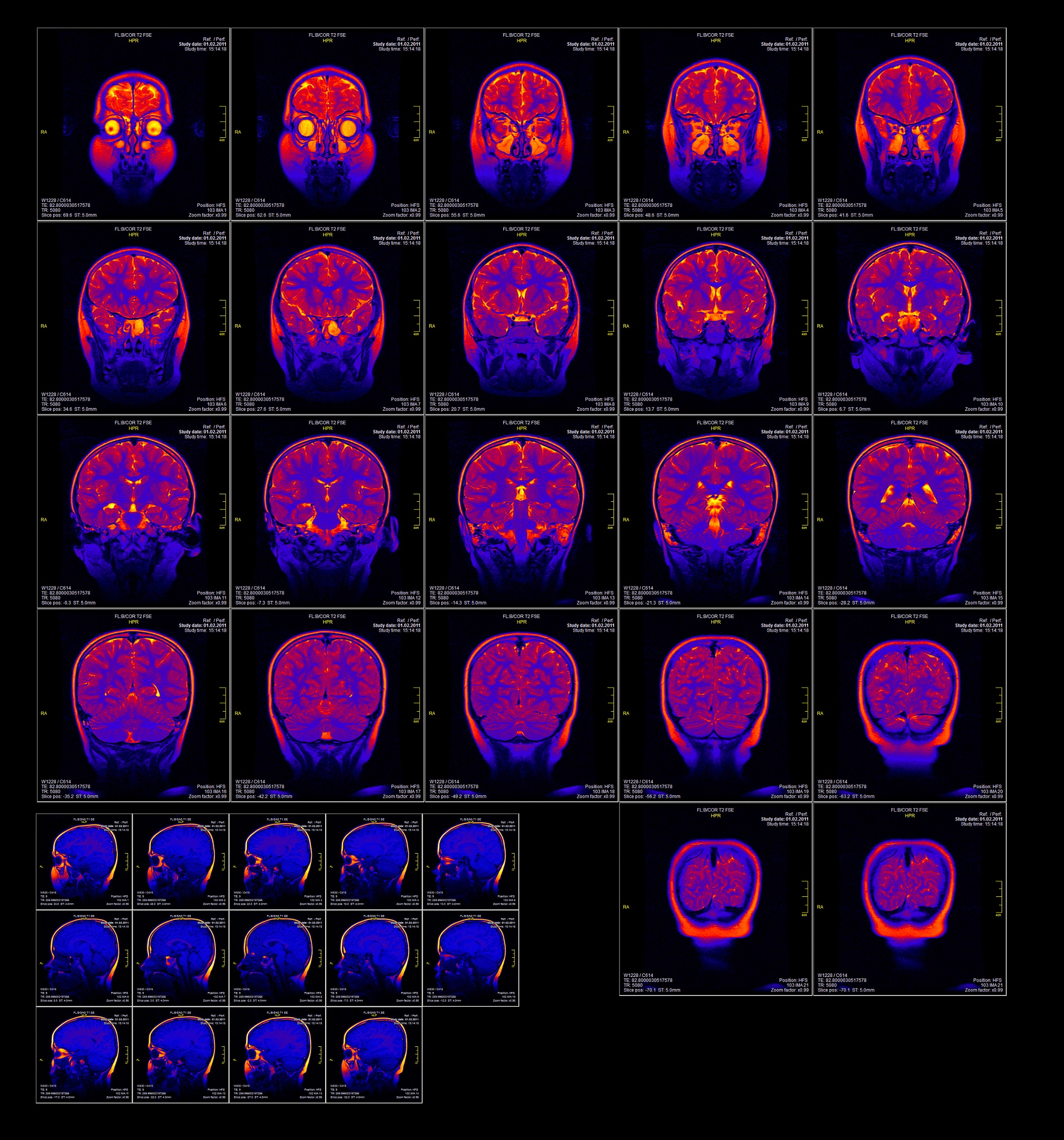
Magnetic resonance imaging
What is magnetic resonance imaging (MRI)? MRI is a used to visualise the structure of the brain and other regions of the body. It uses the magnetic properties inside cells (such as protons) to create a 3D image of the target region. Understanding structural brain alterations in people with schizophrenia may help understand changes in brain development associated with the illness onset or progression, and may help to inform future treatment strategies. What is the evidence for MRI brain structure? Moderate to high quality evidence found grey matter reductions in bilateral frontal lobe, anterior and posterior cingulate gyri, superior and…
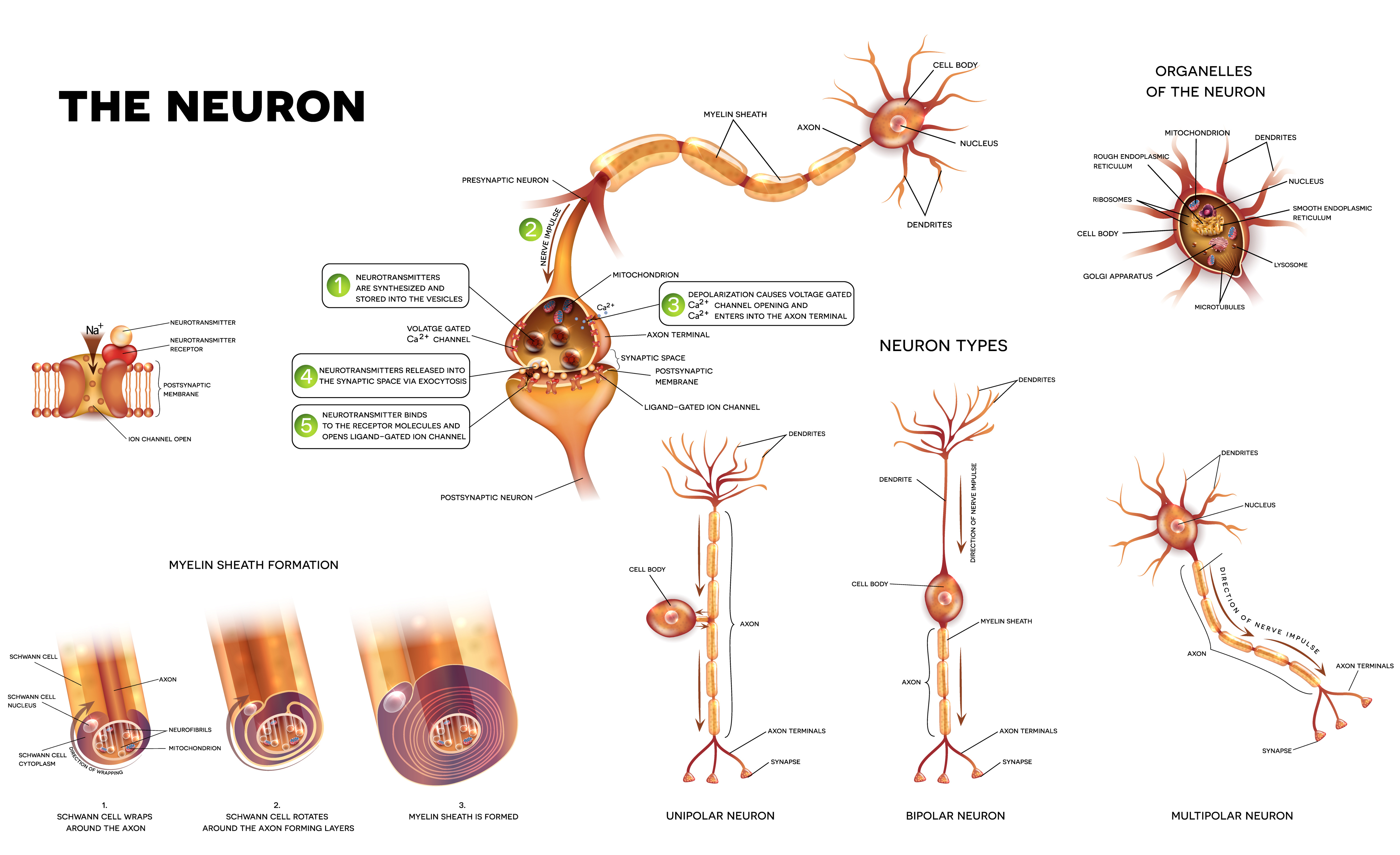
Neuronal changes
What is a neuron? Most neurons have a cell body, an axon, and dendrites. Neurons communicate with other cells over synapses, or gaps between the neurons. Usually, axons send out signals and dendrites receive signals across the synapse, although synapses can also connect an axon to another axon or a dendrite to another dendrite. This process is partly electrical and partly chemical and can be excitatory or inhibitory. A group of connected neurons is called a neural circuit, involving sensory, motor, and interneurons. Studies have shown grey matter reductions in people with schizophrenia, which may involve a loss of neurons…

Optical coherence tomography
What is optical coherence tomography (OCT)? OCT is an imaging technology that assesses the thickness of the peripapillary retinal nerve fibre layer, macular thickness, and volume. It has been used to assess neurologic diseases such as multiple sclerosis, Alzheimer’s disease, and Parkinson’s disease, and more recently, schizophrenia. What is the evidence for OCT? Moderate quality evidence finds a medium-sized effect of thinner overall peripapillary retinal nerve fibre layer thickness in people with schizophrenia compared to controls. There were small effects of thinner nasal and temporal peripapillary retinal nerve fibre layers as well as thinner ganglion cell + inner plexiform layers…
Green - Topic summary is available.
Orange - Topic summary is being compiled.
Red - Topic summary has no current systematic review available.