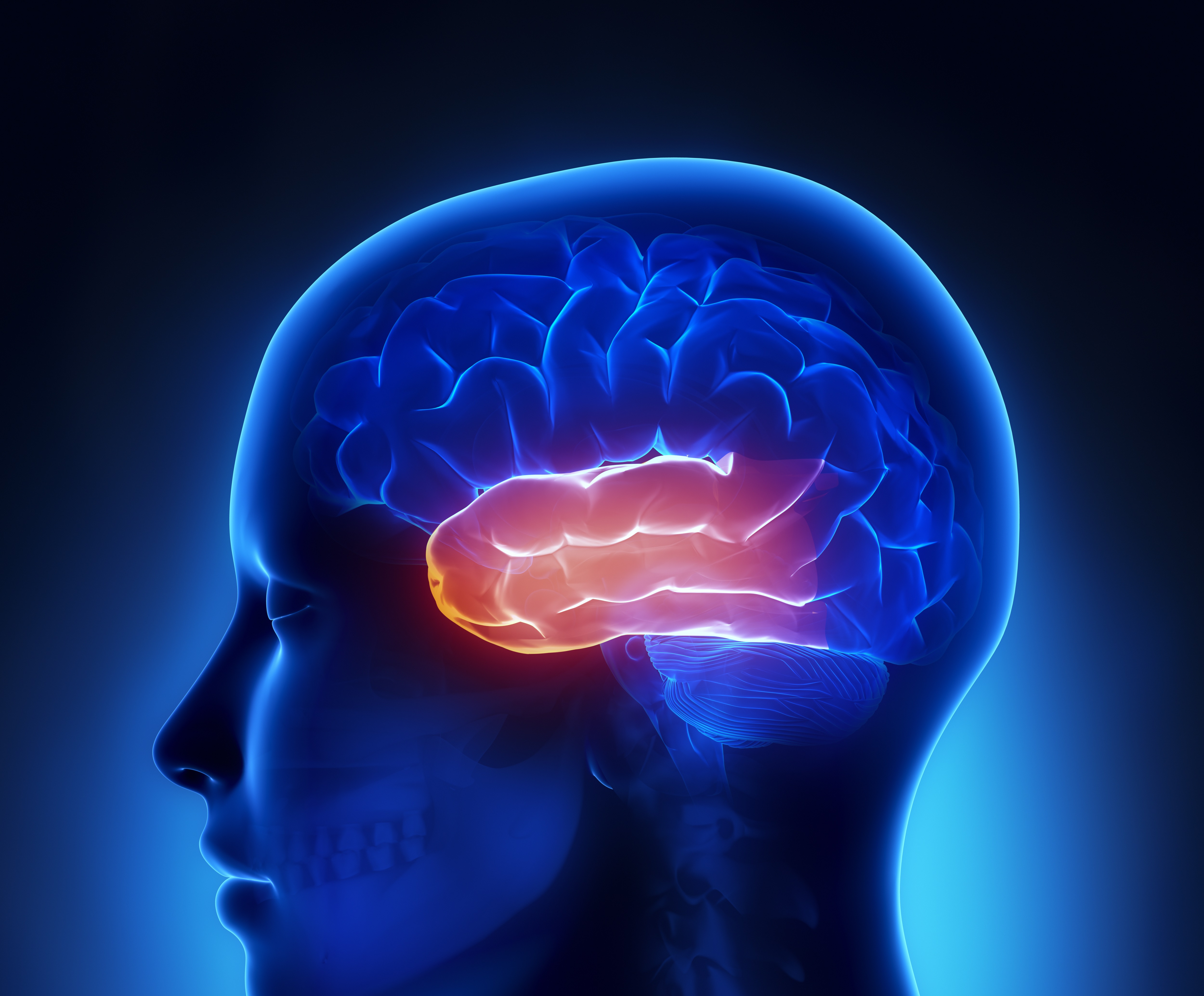
Temporal lobe
What is the temporal lobe?
The temporal lobe is structurally divided into the superior, middle, inferior and medial gyri. The superior temporal gyrus comprises the primary auditory cortex, while nearby temporal regions function in higher level auditory processing, including speech and language. Inferior temporal regions are involved in higher level visual processing and the temporal-occipital gyrus is involved in face processing. The medial temporal lobe comprises the hippocampus and is thought be involved in the formation and propagation of memory.
What is the evidence for temporal lobe alterations?
Structural changes
Moderate to high quality evidence found reduced grey and white matter in the temporal lobe of people with schizophrenia, particularly in the superior temporal gyrus, medial and middle temporal gyrus, and the occipito-temporal gyrus. There was also an absence of normal leftward asymmetry in the planum temporale and excess rightward asymmetry in the superior temporal gyrus (particularly posterior). The severity of auditory hallucinations was associated with grey matter volume reductions in the left superior temporal gyri, (including the rolandic operculum and Heschl’s gyri), and a trend effect for the right superior temporal gyri (including the medial temporal gyrus and Heschl’s gyri).
People with first-episode schizophrenia showed the greatest reductions in the superior and inferior temporal and transverse gyri. There were common decreases in grey matter volume in the left superior temporal gyrus of antipsychotic-naïve and treated first-episode patients compared to controls. Grey matter in the left middle temporal gyrus was increased in antipsychotic-naive patients compared to controls but decreased in treated patients compared to controls.
People at high risk of schizophrenia (relatives or people showing subclinical symptoms) had decreases in the right superior temporal gyrus compared to controls. Relatives also showed reductions in the left inferior temporal gyrus when compared to controls. Relatives showed greater right superior temporal reductions than people with subclinical symptoms, who showed increases in the left superior temporal gyrus compared to controls. People at risk of schizophrenia who developed a psychotic episode showed greater decreases in the right superior temporal gyrus than people at risk who did not develop psychosis.
Functional changes
Moderate quality evidence found increased activation in the superior and middle temporal gyri during auditory hallucinations and decreased activation in the superior temporal gyrus during auditory stimulation in patients compared to controls.
There was increased activation in; the left middle temporal gyrus during episodic memory encoding; the medial temporal gyrus during episodic memory retrieval; the superior temporal and medial temporal gyri during working memory tasks; the right superior temporal gyrus during executive functioning tasks; and the right middle temporal gyrus during timing tasks. There was decreased activation in; the medial temporo-occipital gyrus during memory retrieval tasks; in the superior temporal gyrus of people with schizophrenia during reward anticipation tasks; and lateral and middle temporal regions during linguistic (mostly semantic reading) and theory of mind tasks.
Moderate to high quality evidence found first-degree relatives compared to controls showed increased activation in the right posterior and anterior superior temporal gyrus during cognitive tasks. Moderate to low quality evidence also found increased activation in the right middle temporal gyrus of relatives during emotion tasks. There was decreased activation in schizophrenia compared to autism in bilateral superior temporal gyri during face emotion recognition.
Moderate to high quality evidence finds a medium-sized increase in phosphodiesters in the temporal lobe of people with schizophrenia, with no differences in phosphomonoesters levels. Moderate to low quality evidence found reduced phosphomonoesters and increased phosphodiesters levels in the temporal lobe of people with first-episode psychosis. Moderate to high quality evidence also found decreased N-acetyl aspartate in the temporal lobe of people with first-episode or chronic schizophrenia. There may also be reduced N-acetyl aspartate in people at high-risk of schizophrenia. Moderate quality evidence found reduced translocator protein in the temporal lobe of people with schizophrenia.
October 2020
Fact Sheet Technical Commentary
Green - Topic summary is available.
Orange - Topic summary is being compiled.
Red - Topic summary has no current systematic review available.