
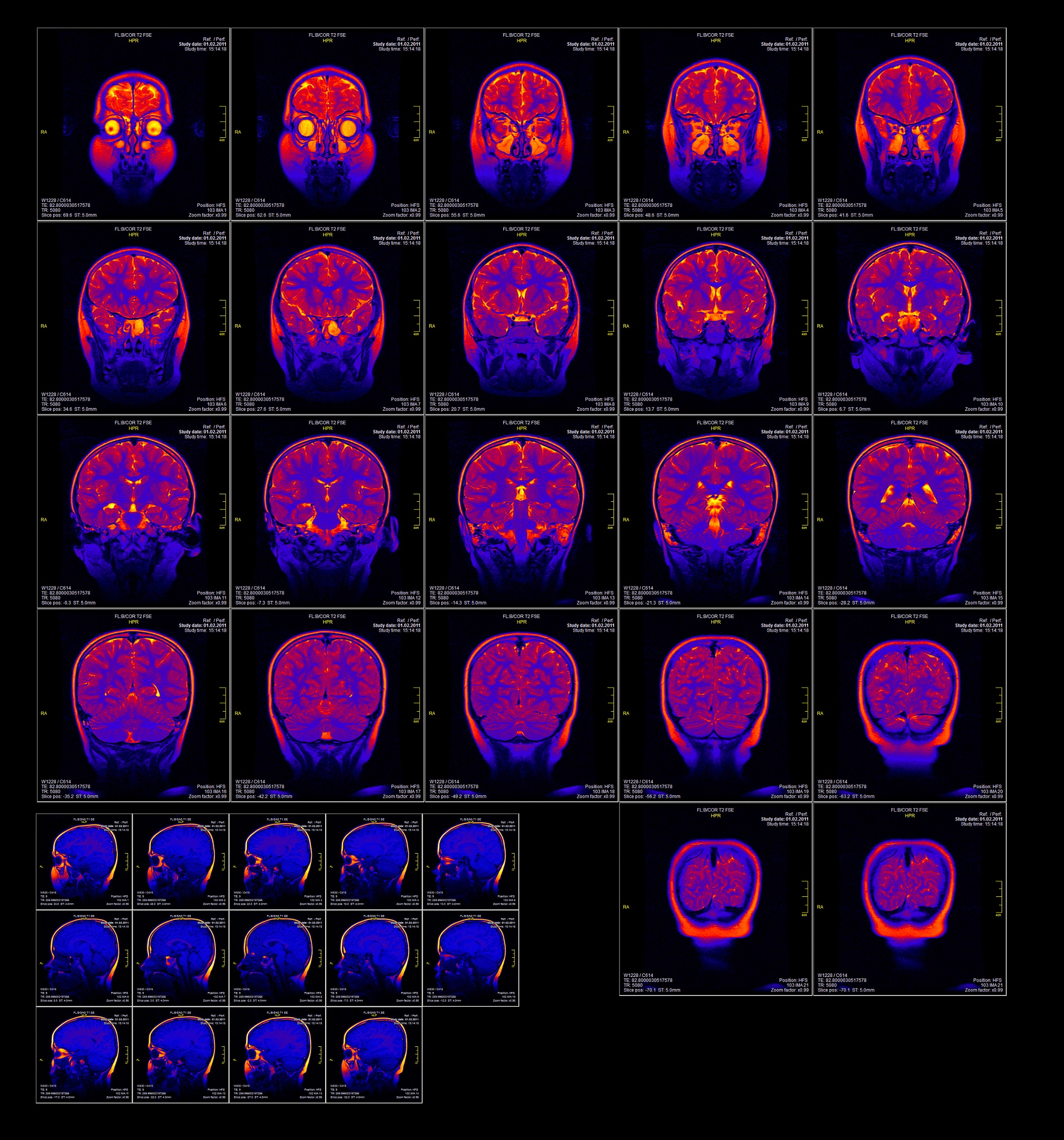
Cerebral blood flow and metabolism
This category provides information regarding changes to cerebral blood flow and metabolism in schizophrenia. These are assessed using functional magnetic resonance imaging (fMRI), magnetic resonance spectroscopy (MRS), positron emission tomography (PET), and single-photon emission computed tomography (SPECT). Click on the links or the tabs below to access the information, or browse via the drop-down menu on the left.
Image: ©2018 Andrew Ostrovsky – stock.adobe.com
Functional magnetic resonance imaging
What is fMRI? Functional magnetic resonance imaging (fMRI) is used to determine functional activation of brain regions when an individual performs tasks (or rests) inside an MRI scanner. Most commonly fMRI studies use visual, auditory, motor or sensory stimuli to evoke neural responses in the brain. Recent fMRI studies also examine activity of the brain at rest. Changes in blood flow are interpreted to represent brain activation (or deactivation) associated with a particular brain state (i.e, while performing a particular activity, or while the brain is at rest). Functional activity has been investigated in people with schizophrenia compared to people…
Magnetic resonance spectroscopy
What is magnetic resonance spectroscopy (MRS)? MRS is a specialised imaging technique that utilises magnetic resonance imaging to investigate biochemical alterations within tissues. Two notable methods of MRS are 1H-MRS (proton-MRS) and 31P-MRS (phosphorus-MRS). Each technique is sensitive to different metabolic compounds. 1H-MRS can be used to measure N-acetylaspartate, an amino acid that is used as a marker of neuronal viability. Decreased levels are associated with neuron death or dysfunction. 1H-MRS is also used to measure creatine, a compound involved in energy metabolism, glutamate, a neurotransmitter, and glutamine, a metabolite of glutamate. 31P-MRS is used to measure phospholipid levels, such…

Positron emission tomography
What is positron emission tomography? Positron emission tomography (PET) is a nuclear based imaging technique that utilises a radioactive tracer to visualise functional brain activity. PET imaging is frequently used in combination with anatomical imaging such as computed tomography (CT) or structural magnetic resonance imaging (MRI). The radioisotopes tracers are coupled with a biological molecule such as glucose, which is used during cellular metabolism and can be used to highlight areas with changes in metabolic activity. Using PET, functional brain activity has been investigated in people with schizophrenia compared to people without schizophrenia to identify regions of increased or decreased…
Single photon emission computed tomography
What is SPECT? Single-photon emission computed tomography (SPECT) is a nuclear based imaging technique that uses radioactive tracers to visualise functional brain activity. SPECT imaging is frequently used in combination with anatomical imaging such as computed tomography (CT) or structural magnetic resonance imaging (MRI). The radioisotope tracers are coupled with a biological molecule such as glucose, which is used during cellular metabolism and can be used to highlight areas with changes in metabolic activity. Functional brain activity has been investigated in people with schizophrenia compared to people without schizophrenia to identify regions of increased or decreased metabolic function or blood…
NeuRA Libraries
-
Bipolar Disorders Library
- Diagnosis and assessment
-
Signs and symptoms
- General signs and symptoms
-
Cognition
- Attention
- Cognition and bipolar disorder symptoms
- Cognition and bipolar disorder type
- Cognition and functioning
- Cognition in bipolar disorder and major depression
- Cognition in bipolar disorder and schizophrenia
- Cognition in children with bipolar disorder
- Cognition in first-episode bipolar disorder
- Cognition in late-life bipolar disorder
- Cognition in relatives
- Decision making
- Episodic future thinking
- Executive functioning
- Insight
- IQ and global cognition
- Language
- Learning
- Memory
- Metacognition
- Processing speed
- Reasoning and problem solving
- Social cognition
- Visuospatial ability
-
Treatments
-
Physical
- Non-pharmaceutical
-
Pharmaceutical
- Mood stabilisers
- Antidepressants
- Antipsychotics
- Adjunctive and alternative treatments
-
Medications for specific symptoms and populations
- Medication during pregnancy and breastfeeding
- Medication for aggression and agitation
- Medication for bipolar II disorder
- Medication for bipolar versus unipolar depression
- Medication for children
- Medication for cognitive symptoms
- Medication for dual diagnosis
- Medication for elderly people
- Medication for first-episode bipolar disorder
- Medication for high-risk groups
- Medication for mixed states
- Medication for rapid cycling
- Medication for relapse prevention
- Medication for suicide and self-harm prevention
- Medication for treatment resistance
- Other pharmaceutical topics
-
Psychotherapy
- Therapies
-
Therapies for specific symptoms and populations
- Therapies for children
- Therapies for cognition
- Therapies for dual diagnosis
- Therapies for first-episode bipolar disorder
- Therapies for high-risk groups
- Therapies for internalised stigma
- Therapies for parents with bipolar disorder
- Therapies for trauma-related symptoms
- Therapies for treatment non-adherence
- Therapies for weight gain
-
Physical
-
Risk factors and antecedents
- Antecedents
-
Environmental risk factors
- Adult life events
- Childbirth
- Childhood adversity
- Environmental toxins
- Ethnicity
- Familial factors
- Infectious agents
- Maternal diet during pregnancy
- Maternal illness during pregnancy
- Migration
- Obstetric complications
- Parental age at birth
- Parental education
- Parental psychological factors
- Rheumatoid arthritis
- Sibship
- Socioeconomic status
- Substance use
- Traumatic brain injury
- Urbanicity
- Winter birth
-
Illness course and outcomes
- Absconding
- Age at onset
- Attitudes to medication
- Creativity
- Criminal offending, aggression and violence
- Criminal victimisation
- Cultural differences
- Dietary intake
- Drug and alcohol use
- Employment
- First-episode bipolar disorder
- Functional outcomes
- Homelessness
- Hope
- Menopause
- Mortality
- Parenthood
- Pathways to care
- Pediatric bipolar disorder
- Physical activity
- Physical health monitoring
- Quality of care
- Quality of life
- Recovery
- Relapse
- Relationships
- Religiosity
- Smoking
- Stigma
- Suicide and self-harm
- Treatment non-adherence
- Treatment resistance
- Insights for families
-
Physical features
-
Functional changes
- Body functioning
-
Biochemical changes
- Brain pH and lactate
- Calcium
- Complex I and IV
- Dopamine
- GABA
- Gut microbiota
- Homocysteine
- Hypothalamic-pituitary-adrenal axis
- Infectious agents
- Inflammation and immune system dysfunction
- Insulin-like growth factor
- Lipids
- Neurometabolites
- Neuropeptides
- Neurotrophins
- NMDA receptor function
- Oxidative stress
- S100 proteins
- Serotonin
- Vitamin B
- Cerebral blood flow and metabolism
- Electrophysiology
- Structural changes
-
Functional changes
-
Co-occurring conditions
- Mental disorders
-
Physical disorders
- Asthma
- Autoimmune disease
- Blood disorders
- Cancer
- Cerebrovascular disease
- Dementia
- Diabetes
- Digestive disorders
- Epilepsy
- Fibromyalgia
- Heart disease
- Infectious diseases
- Metabolic syndrome
- Musculoskeletal and connective tissues
- Obesity
- Optical alterations
- Osteoporosis
- Pain and migraine
- Parkinson’s disease
- Peripheral vascular disease
- Premenstrual syndrome
- Respiratory disease
- Skin disorders
- Sleep apnea
- Thyroid disorders
- Venous thromboembolism
- Visual impairment
- Wilson’s disease
- Substance use
-
Incidence and prevalence
- General
- Incidence
-
Prevalence
- Prevalence in children
- Prevalence in elderly people
- Prevalence in forensic settings
- Prevalence in homeless populations
- Prevalence in indigenous populations
- Prevalence in males vs. females
- Prevalence in migrants
- Prevalence in primary care settings
- Prevalence in problem gamblers
- Prevalence in veterans
- Spatial variation in prevalence
- Worldwide prevalence
- General information
-
Post-Traumatic Stress Disorder Library
- Diagnosis and assessment
- Signs and symptoms
-
Treatments
- Physical
-
Psychotherapy
-
Therapies
- Acceptance and commitment therapy
- All psychological therapies for PTSD
- Animal-assisted psychotherapy
- Brief eclectic psychotherapy
- Cognitive behavioural therapy
- Cognitive therapies
- Creative arts therapy
- Emotion-focussed therapies
- Emotional freedom techniques
- Exposure therapies
- Expressive writing
- Eye movement desensitization and reprocessing
- Family therapies
- Group therapies
- Hypnotherapy
- ICU diaries
- Imagery rehearsal therapy
- Interpersonal psychotherapy
- Memory specificity training
- Metacognitive therapy
- Mindfulness and meditation
- Narrative exposure therapy
- Present-centred therapy
- Psychoeducation
- Self-help treatment
- Stress inoculation training
- Stress management
- Structured approach therapy
- Supportive counselling
- Telemental health
- Trauma affect regulation
-
Therapies for specific symptoms and populations
- Therapies for adults with a history of childhood abuse
- Therapies for children and adolescents
- Therapies for complex PTSD
- Therapies for dual diagnosis
- Therapies for people living in low- and middle-income countries
- Therapies for prevention of PTSD
- Therapies for refugees and asylum seekers
- Therapies for soldiers and veterans
-
Therapies
-
Risk factors
- Personal characteristics
-
Trauma characteristics
- Abuse and violence
- Bereavement
- Direct vs. indirect exposure
- Disasters
- Epidemics and pandemics
- Imprisonment
- Intentional vs. unintentional traumas
- Interpersonal vs. non-interpersonal traumas
- Migration and displacement
- Moral injury
- Physical injury and illness
- Pregnancy and childbirth
- Road traffic accidents
- Time post-trauma
- Trauma severity
- War and terrorism
- Illness course and outcomes
- Insights for families
- Physical features
- Co-occurring conditions
-
Incidence and prevalence
-
Incidence
- Incidence in abuse and violence survivors
- Incidence in bereaved people
- Incidence in caregivers
- Incidence in children and adolescents
- Incidence in criminal offenders
- Incidence in disaster survivors
- Incidence in elderly people
- Incidence in epidemic and pandemic survivors
- Incidence in firefighters
- Incidence in healthcare workers
- Incidence in homeless populations
- Incidence in humanitarians
- Incidence in indigenous people
- Incidence in journalists
- Incidence in jurors
- Incidence in males vs. females
- Incidence in medical patients
- Incidence in police officers
- Incidence in problem gamblers
- Incidence in psychiatric inpatients
- Incidence in public transport drivers
- Incidence in refugees and asylum seekers
- Incidence in rescue teams
- Incidence in road traffic accident survivors
- Incidence in soldiers and veterans
- Incidence in war and terrorism survivors
- Worldwide incidence
-
Prevalence
- Prevalence in abuse and violence survivors
- Prevalence in bereaved people
- Prevalence in caregivers
- Prevalence in children and adolescents
- Prevalence in disaster survivors
- Prevalence in elderly people
- Prevalence in epidemic and pandemic survivors
- Prevalence in firefighters
- Prevalence in forensic settings
- Prevalence in healthcare workers
- Prevalence in homeless populations
- Prevalence in humanitarians
- Prevalence in indigenous people
- Prevalence in journalists
- Prevalence in jurors
- Prevalence in males vs. females
- Prevalence in medical patients
- Prevalence in police officers
- Prevalence in problem gamblers
- Prevalence in psychiatric patients
- Prevalence in refugees
- Prevalence in rescue teams
- Prevalence in road traffic accident survivors
- Prevalence in soldiers and veterans
- Prevalence in train drivers
- Prevalence in war and terrorism survivors
- Spatial variation in prevalence
- Worldwide prevalence
-
Incidence
- General information
-
Schizophrenia Library
- Diagnosis and assessment
-
Signs and symptoms
-
General signs and symptoms
- Attachment styles
- Dermatoglyphics
- Disorganised symptoms
- Dissociation
- Early detection
- Functional laterality
- Minor physical anomalies
- Morphometrics
- Movement disorders
- Negative symptoms
- Neurological soft signs
- Olfactory functioning
- Pain sensitivity
- Personality and temperament
- Positive symptoms
- Psychotic relapse
- Sleep disturbance
- Temperature regulation
-
Cognition
- Attention
- Cognition in high-risk groups
- Cognition in schizophrenia and bipolar disorder
- Cognitive functioning related to symptoms
- Decision making
- Defeatist performance beliefs
- Episodic future thinking
- Executive functioning
- Information processing
- Insight
- IQ and global cognition
- Language
- Learning
- Memory
- Metacognition
- Psychomotor ability
- Reasoning and problem solving
- Rigidity
- Social cognition
- Time perception
- Tone perception
- Visuospatial ability
- Voice patterns
-
General signs and symptoms
-
Treatments
-
Physical
- Non-pharmaceutical
-
Pharmaceutical
-
First-generation antipsychotics
- All antipsychotics versus placebo
- Benperidol
- Bromperidol
- Chlorpromazine
- Droperidol
- First versus second generation
- Flupentixol
- Fluphenazine
- Fluspirilene
- Haloperidol
- Levomepromazine
- Loxapine
- Metiapine
- Molindone
- Penfluridol
- Perazine
- Perphenazine
- Pimozide
- Piperacetazine
- Pipotiazine
- Sulpiride
- Thioridazine
- Trifluoperazine
- Zuclopenthixol
-
Second-generation antipsychotics
- All antipsychotics versus placebo
- Amisulpride
- Aripiprazole
- Asenapine
- Blonanserin
- Brexpiprazole
- Cariprazine
- Carpipramine
- Clocapramine
- Clotiapine
- Clozapine
- First versus second generation
- Iloperidone
- Lurasidone
- Mosapramine
- Olanzapine
- Paliperidone
- Perospirone
- Quetiapine
- Remoxipride
- Risperidone
- Second versus second generation
- Sertindole
- Ziprasidone
- Zotepine
-
Adjunctive medications
- Adenosine modulators
- Amphetamines
- Analeptics
- Anti-inflammatory
- Anticholinergic
- Anticonvulsants
- Anticraving agents
- Antidepressants
- Benzodiazepines
- Beta blockers
- Calcium channel blockers
- Catecholamines
- Cholinergic medications
- Cholinesterase inhibitors
- Erythropoietin
- Essential fatty acids
- Estrogen
- GABA agonists
- GHB
- Glutamate receptor modulators
- Herbal medicines
- Medicinal cannabis
- Mood stabilisers
- Oxytocin
- Promethazine
- Serotonin modulators
- Sodium nitroprusside
- Statins
- Testosterone
- Alternative medications
-
Medications for specific symptoms and populations
- Medications during pregnancy and breastfeeding
- Medications for aggression and agitation
- Medications for best adherence
- Medications for childhood and early-onset schizophrenia
- Medications for cognitive symptoms
- Medications for constipation
- Medications for depressive symptoms
- Medications for dual diagnosis
- Medications for elderly people and people with late-onset schizophrenia
- Medications for first-episode psychosis
- Medications for hyperprolactinemia
- Medications for hypersalivation
- Medications for movement disorders
- Medications for negative symptoms
- Medications for relapse prevention
- Medications for schizoaffective disorder
- Medications for sexual dysfunction
- Medications for sleep disturbance
- Medications for smoking cessation
- Medications for social functioning
- Medications for treatment-resistant schizophrenia
- Medications for weight gain
- Therapies and medications for high-risk groups
- Other
-
Side effects
- Blood disorders
- Bone density
- Cancer
- Cardiometabolic changes and weight gain
- Constipation
- Dysphagia
- Extrapyramidal
- Hyperprolactinaemia
- Hypersalivation
- Hyponatraemia
- Mortality
- Neuroleptic malignant syndrome
- Neutropenia
- Oculogyric crisis
- Pancreatitis
- Polycystic ovarian syndrome
- Sedation
- Seizures
- Sexual dysfunction
- Thyroid dysfunction
-
First-generation antipsychotics
-
Psychotherapy
-
Therapies
- Acceptance and commitment therapy
- Animal-assisted therapy
- Art and drama therapies
- Case management
- Cognitive behavioural therapy
- Community care
- Cost
- Crisis intervention
- Crisis planning
- Dance therapy
- Day centres and day hospitals
- Dialectical behavioural therapy
- Distraction techniques
- Educational therapies
- Enriched intervention
- Family intervention
- Genetics counselling
- Group therapy
- Home-based care
- Hypnosis
- Inpatient and outpatient care
- Integrated care
- Life skills programs
- Metacognitive training
- Mindfulness
- Monetary incentives
- Morita therapy
- Music therapy
- Nidotherapy
- Open dialogue approach
- Peer support
- Physical restraint
- Prevention programs
- Problem solving skills training
- Psychodynamic psychotherapy
- Shared decision making
- Strengths-based delivery
- Supported housing
- Supportive therapy
- Telemental health
- Therapeutic relationship
- Token economies
- Trauma-focused therapies
- User-held records
- Virtual reality and avatar therapy
-
Therapies for specific symptoms and populations
- Therapies and medications for high-risk groups
- Therapies for childhood onset and early onset schizophrenia
- Therapies for cognition
- Therapies for dual diagnosis
- Therapies for fathers with schizophrenia
- Therapies for first-episode psychosis
- Therapies for insight
- Therapies for internalised stigma
- Therapies for mothers with schizophrenia
- Therapies for negative symptoms
- Therapies for positive symptoms
- Therapies for PTSD symptoms
- Therapies for quality of life
- Therapies for schizoaffective disorder
- Therapies for smoking
- Therapies for social functioning
- Therapies for treatment non-adherence
- Therapies for treatment resistance
- Therapies for unemployment
- Therapies for weight gain
-
Therapies
-
Physical
-
Risk factors and antecedents
-
Antecedents
- Autonomic nervous system anomalies
- Behavioural disturbances and psychopathology
- Dermatoglyphic anomalies
- Eye tracking anomalies
- Face emotion processing anomalies
- Height and body mass index
- IQ and academic performance
- Minor physical anomalies
- Motor dysfunction
- Olfactory identification deficits
- Sleep disturbance
- Speech and hearing deficits
- Stress responsivity anomalies
-
Environmental risk factors
- Adult life events
- Childhood adversity
- Environmental toxins
- Ethnicity
- Family relationships
- Famine
- Genetic and non-genetic risk
- Infectious agents
- Latitude, climate and winter birth
- Marital status
- Maternal diet and body mass index
- Maternal illness during pregnancy
- Migration
- Obstetric complications
- Parental age at birth
- Parental education
- Parental psychological factors
- Sex differences
- Sibship
- Social capital
- Socioeconomic status
- Substance use
- Traumatic brain injury
- Urban environment
- Genetic risk factors
-
Antecedents
-
Illness course and outcomes
- Absconding
- Age at onset
- Childhood and early-onset schizophrenia
- Creativity
- Criminal offending, aggression and violence
- Criminal victimisation
- Cultural differences
- Diet
- Drug and alcohol use
- Duration of untreated psychosis
- Duration of untreated psychosis and outcomes
- Electronic device use
- Employment
- First-episode psychosis
- Functional outcomes
- Homelessness
- Hope
- Late-onset schizophrenia
- Loneliness
- Mortality
- Parenthood
- Pathways to care
- Physical activity
- Physical health monitoring
- Psychotic relapse
- Quality of care
- Quality of life
- Relationships
- Religiosity
- Remission and recovery
- Sex differences
- Smoking
- Stigma and attitudes towards mental health
- Suicide and self-harm
- Treatment adherence
- Treatment-resistance
- Insights for families
-
Physical features
-
Functional changes
- Body functioning
-
Biochemical changes
- Brain pH and lactate
- cAMP
- Cholesterol
- Dopamine
- Endocannabinoids
- GABA
- Hormonal changes
- Hypothalamic-pituitary-adrenal axis
- Infectious agents
- Inflammation and the immune system
- Lipids
- Neurometabolites
- Neurotrophins
- Nitric oxide
- NMDA receptor function
- Oxidative stress
- S100 Proteins
- Serotonin
- Synaptic proteins
- Trace elements
- Vascular endothelial growth factor
- Vitamin B
- Vitamin D
- Cerebral blood flow and metabolism
- Electrophysiology
- Structural changes
- Brain regions
-
Functional changes
-
Co-occurring conditions
- Mental disorders
-
Physical disorders
- Auditory system dysfunction
- Autoimmune diseases
- Blood disorders
- Cancer
- Cerebrovascular disease
- Dementia
- Dental disease
- Diabetes
- Digestive disorders
- Epilepsy
- Heart disease
- Infectious diseases
- Metabolic syndrome
- Musculoskeletal and connective tissues
- Obesity
- Optical alterations
- Peripheral vascular disease
- Polycistic ovary syndrome
- Postoperative complications
- Reproductive and urological disorders
- Respiratory system dysfunction
- Sexual dysfunction
- Skin disorders
- Sleep apnea
- Thyroid disorders
- Underweight
- Venous thromboembolism
- Substance use
-
Incidence and prevalence
- General
- Incidence
-
Prevalence
- Prevalence in children
- Prevalence in elderly people
- Prevalence in forensic settings
- Prevalence in homeless populations
- Prevalence in indigenous populations
- Prevalence in inpatients
- Prevalence in males vs. females
- Prevalence in migrants
- Prevalence in refugees
- Prevalence in veterans and conflict settings
- Spatial variation in prevalence
- Worldwide prevalence
- General information
-
Podcast Library
Green - Topic summary is available.
Orange - Topic summary is being compiled.
Red - Topic summary has no current systematic review available.