Cingulate cortex
What is the cingulate cortex?
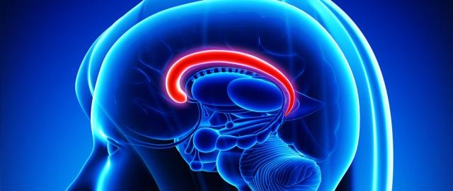
The cingulate cortex is part of the medial frontal cortex, located immediately dorsal to the corpus callosum along the sagittal midline. The anterior cingulate cortex has three key divisions which may be functionally distinct (dorsal, rostral, subcallosal). The dorsal part of the anterior cingulate cortex has reciprocal connections with the prefrontal and parietal cortices as well as the frontal eye fields, and plays a primary role in balancing top-down and bottom-up processing of external stimuli; that is, monitoring behaviour and incoming stimuli in the context of current goals, and assigning control to other areas in the brain when required. By contrast, the ventral (rostral and subcallosal) parts of the anterior cingulate cortex are connected with amygdala, nucleus accumbens, hypothalamus, and insula, and are implicated in assessing the salience of sensory information, and regulating emotion and autonomic activity.
What is the evidence for cingulate cortex anomalies?
Structural changes
Moderate to high quality evidence found reductions in grey and white matter in bilateral regions of the cingulate cortex of people with schizophrenia. Specifically, there were grey matter reductions in the anterior and posterior cingulate gyrus of people with schizophrenia and reduced grey matter in the left anterior cingulate/paracingulate gyrus and right dorsal anterior cingulate of first-episode patients. High-risk individuals showed decreases in bilateral median cingulate and the right anterior cingulate gyrus compared to controls and increases in the left anterior cingulate compared to first-episode patients.
Functional changes
Moderate quality evidence found decreased activity in the posterior cingulate cortex of people with schizophrenia at rest compared to controls. There was under-activation in the anterior and middle cingulate cortex of people with schizophrenia during attention and inhibition tasks, and over-activation in the anterior cingulate cortex during working memory tasks. During executive functioning, there was increased activity in the left cingulate gyrus and decreased activity in right cingulate gyrus. During cognitive control, there was decreased activity in the bilateral anterior cingulate/paracingulate gyrus. During auditory stimulation, there was decreased activation in the anterior cingulate cortex, and during theory of mind tasks, there was decreased activity in the left cingulate gyrus. During reward anticipation tasks there was reduced activation in the right median cingulate/paracingulate gyri. In relatives of people with schizophrenia, there was decreased activity in the left cingulate gyrus during executive functioning and working memory tasks compared to controls.
Moderate quality evidence found decreased N-acetylaspartate and N-acetylaspartate/creatine ratio in the anterior cingulate gyrus of people with schizophrenia and their first-degree relatives. High quality evidence finds a small decrease in glutathione in the anterior cingulate of people with schizophrenia.
Structural and functional changes
Moderate to high quality evidence found decreased grey matter volume and decreased functional activity in the left medial posterior cingulate/paracingulate gyrus in drug-free, first-episode patients. Moderate quality evidence found decreased grey matter volume and decreased functional activation in the right medial frontal/anterior cingulate, and decreased grey matter volume and increased functional activation in the left medial frontal/anterior cingulate of people with first-episode schizophrenia, regardless of medication status.
Moderate quality evidence finds decreased activation in the bilateral anterior cingulate and left posterior cingulate of people with schizophrenia compared to people with an autism spectrum disorder during facial emotion recognition tasks. During theory of mind tasks, there was increased activation in the left posterior cingulate cortex in people with schizophrenia compared to people with an autism spectrum disorder. Moderate to low quality evidence found similar grey matter volume decreases in the right posterior cingulate cortex of people with schizophrenia and people with an autism spectrum disorder.
October 2020
Fact Sheet Technical Commentary
Green - Topic summary is available.
Orange - Topic summary is being compiled.
Red - Topic summary has no current systematic review available.